Figures & data
Figure 1 Equipment required to perform tube CTT using the modified technique (with airway occlusion).
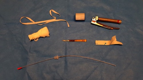
Figure 2 Sagittal section of weaner pig larynx. Blue box indicates thyroid cartilage. Red box indicates cross-section of ventral cricoid cartilage and black arrow indicates the position of the cricothyroid notch.
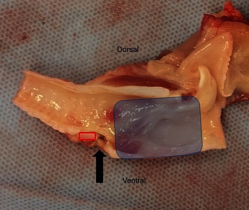
Figure 3 The CTT procedure performed on the pig model (A-G).
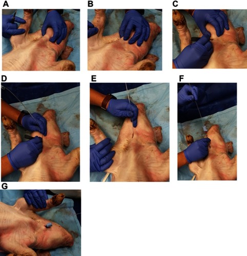
Figure 4 A dissected view and example of a wide approach of the CTT procedure. The cricothyroid ligament is incised (A). Urinary catheter is placed (B). A size 3.5 uncuffed endotracheal tube is placed into the incision (C).
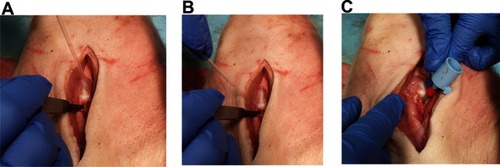
Table 1 Pig characteristics and procedure-related data
Figure 5 Time-course of changes in ETCO2 for the 10 mins following completion of the procedure.
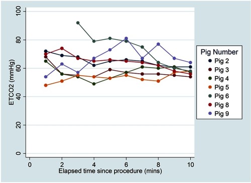
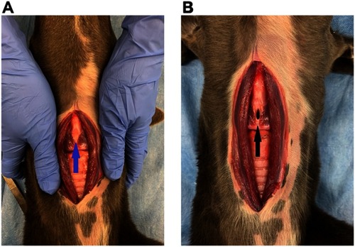
Figure 6 Dissection depicting the ventral laryngeal anatomy in a cadaver dog. (A) Blue arrow points to intact cricothyroid membrane and ligament. (B) Black arrow indicates the incision in the cricothyroid ligament. The cricothyroid membrane is located on the ventral aspect of the larynx, joining the caudoventral border of the thyroid cartilage and the cranioventral aspect of the cricoid cartilage. The medial part of the cricothyroid membrane is termed the cricothyroid ligament. The ligament is devoid of a major blood supply but may have small vessels associated near the cricoid and thyroid attachments.Citation26