
Figures & data
Figure 1 The normal equine stomach showing the four distinct anatomical regions. A, ventral fundus, B, cardia; C, pylorus; D, dorsal fundus. Image adapted with permission of Equine Veterinary Journal. Martineau H, Thompson H, Taylor D. Pathology of gastritis and gastric ulceration in the horse. Part 1: range of lesions present in 21 mature individuals. Equine Vet J. 2009;41(7):638–644.Citation23
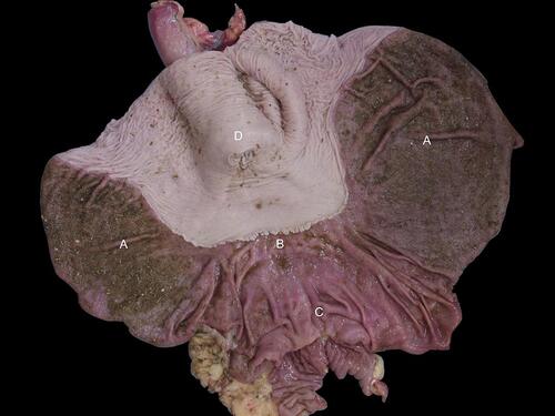
Figure 2 Hyperkeratosis (white arrow). Notice the thickened, corrugated squamous mucosa surrounding the cardia. Regions of hyperkeratosis often have a characteristic yellowish discoloration. Image property of the author.
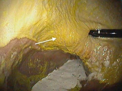
Figure 3 Parakeratotic hyperkeratosis (dashed arrow). Bar = 400 μm. Image reproduced with permission of Equine Veterinary Journal. Martineau H, Thompson H, Taylor D. Pathology of gastritis and gastric ulceration in the horse. Part 1: range of lesions present in 21 mature individuals. Equine Vet J. 2009;41(7):638–644.Citation23
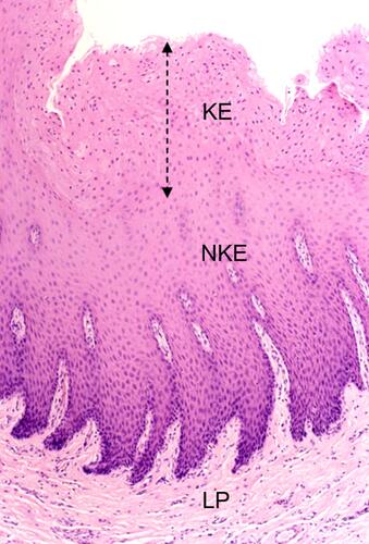
Figure 4 Erosion (white arrow). Notice the focal region of squamous mucosa that is smooth and reddish/pink in appearance. This represents thinning of the squamous epithelium and loss of the keratinized layer. Image courtesy of Henny Martineau.
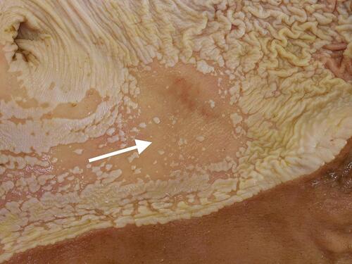
Figure 5 Erosion (black arrow) adjacent to normal squamous epithelium. Bar = 1000 μm. Notice the dramatic thinning of the affected epithelium. Image reproduced with permission of Equine Veterinary Journal. Martineau H, Thompson H, Taylor D. Pathology of gastritis and gastric ulceration in the horse. Part 1: range of lesions present in 21 mature individuals. Equine Vet J. 2009;41(7):638–644.Citation23
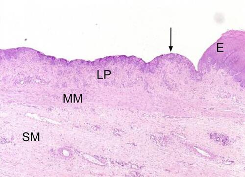
Figure 6 Ulcer (white arrow). Notice the characteristic depressed center surrounded by a prominent margin of tissue. Image property of the author.
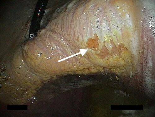
Figure 7 Bleeding ulcer (white arrow). Ulcers may be associated with active hemorrhage when severe. Image property of the author.
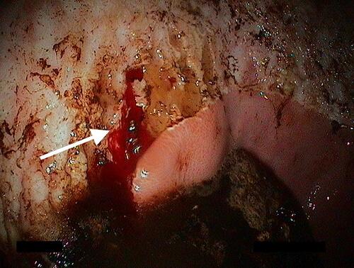
Figure 8 Ulcer (single black arrow) adjacent to normal squamous epithelium. Notice the complete loss of epithelium in the ulcer bed and exposure of the underlying lamina propria. Prominent epithelial projections can be seen in the normal mucosa immediately adjacent to the ulcer (double black arrows). Bar = 1000 μm. Image reproduced with permission of Equine Veterinary Journal. Martineau H, Thompson H, Taylor D. Pathology of gastritis and gastric ulceration in the horse. Part 1: range of lesions present in 21 mature individuals. Equine Vet J. 2009;41(7):638–644.Citation23
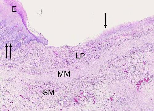
Table 1 Prevalence of ESGD in Different Populations of Horses and Associated Risk Factors
Table 2 Reported Dose Rates for Oral Omeprazole for the Treatment or Prophylaxis of ESGD