Figures & data
Table 1 List and Composition of the Vaccines Used in This Study
Figure 2 Serological response of the gilts over time to vaccination and challenge measured by a commercial blocking ELISA. The dashed line indicates the cut-off of the ELISA.
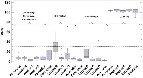
Table 2 Serum Virus Neutralization Titers of the Gilts After Vaccination and Pre-Challenge Against 100 TCID50 PPV1-HUN Strain
Table 3 Physiological Parameters of the Litters
Table 4 Viral Genome Copy Load and Virus Isolation Measurements Results
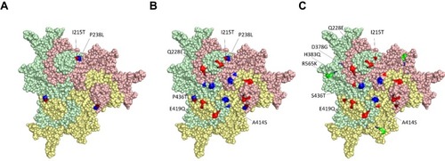
Figure 1 Surface amino acid modifications among different PPV strains. All altered amino acids highlighted on the surface of a capsid trimer (the three inter-linked identical monomers are shown in different colors), but changes only on one chain are annotated. Numbering is indicated according to NADL-2 VP2 positions. (A) K22 vs Kresse. Altered neighbouring surface amino acids highlighted with red and blue. (B) K22 vs 27a. Surface amino acid changes are indicated with red and blue. (C) NADL-2 vs 27a. Surface amino acid changes similar to K22 v. 27a are highlighted with red, other amino acid changes are indicated by blue and green.