
Figures & data
Figure 1 Abdominal radiographs with left, right, and ventrodorsal views depicting decreased serosal detail in the mid-abdomen. No mass effect was visualized.
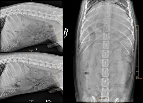
Figure 2 Ultrasound images of enlarged hypoechoic jejunal lymph nodes (A and B) and hypoechoic pancreas, suggesting pancreatitis (C).
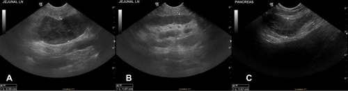
Table 1 Case #1 Salmonella Species Susceptibility Results
Table 2 Case #2 Salmonella Species Susceptibility Results