Figures & data
Figure 1 Fine-needle aspirate cytology of a cranial abdominal mass displaying numerous extracellular and intrahistiocytic Cryptococcus spp. yeasts with prominent nonstaining capsules, narrow-based budding (arrow), and occasional chains of organisms.
Notes: Modified Wright’s stain (Diff-Quik®). 60× objective. Bar = 20 μm.
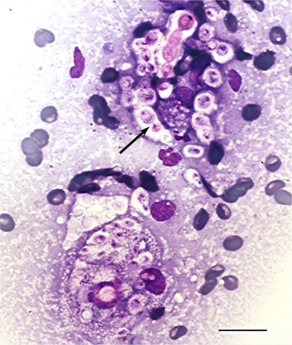
Figure 2 Gross necropsy photograph of intestinal tract with large cryptococcal lesion (demarcated by arrows) adhered to the root of the mesentery.
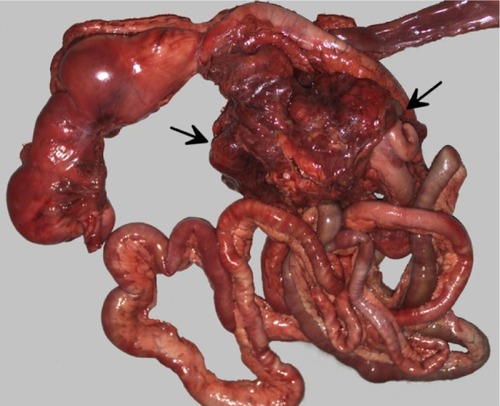
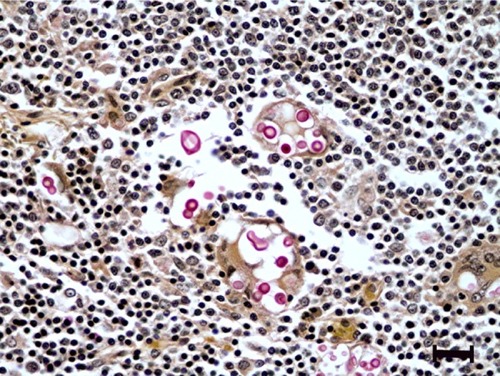
Figure 3 Histologic section of mesenteric lymph node with histiocytic inflammation and extracellular cryptococcal yeast organisms (pink).
Notes: Mucicarmine stain. Bar = 20 μm.