
Figures & data
Figure 1 Measurement of circumferential and radial strain and strain rate.
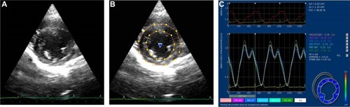
Figure 2 Measurement of longitudinal strain and strain rate.
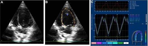
Figure 3 An example of six segments of the left ventricle for measurement of segmental heterogeneity in the circumferential direction using two-dimensional speckle-tracking echocardiography obtained from the right parasternal short-axis view at the level of the papillary muscle.
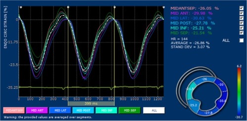
Figure 4 An example of six segments of the left ventricle for measurement of intra-ventricular mechanical synchrony in the longitudinal direction using two-dimensional speckle-tracking echocardiography obtained from the left parasternal apical four-chamber view.
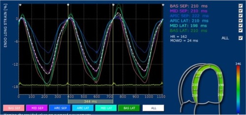
Table 1 The descriptive statistics of two-dimensional and M-mode echocardiographic parameters of 34 clinically healthy cats
Table 2 Segmental and global circumferential, radial, and longitudinal strain and strain rate values determined using two-dimensional speckle-tracking echocardiography of the left ventricle in 34 clinically healthy cats
Table 3 Echocardiographic parameters of left ventricular heterogeneity and synchrony in 34 clinically healthy cats
Table 4 The intra-observer CV values derived from the parameters measured using two-dimensional speckle-tracking echocardiography