Figures & data
Figure 1 Morgellons patient’s lower legs. Similar lesions covered her trunk and arms. There were no excoriations or secondary infections. Photo courtesy of Cindy Casey, Charles E Holman Foundation, Austin, Texas. Reproduced with permission.
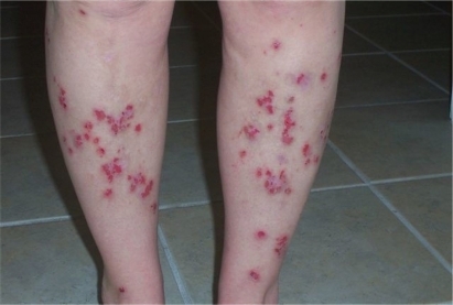
Figure 2 Morgellons patient’s back. Note that lesions and scars occur in areas that could not have been reached by the patient. Photo courtesy of Cindy Casey, Charles E Holman Foundation, Austin, Texas. Reproduced with permission.
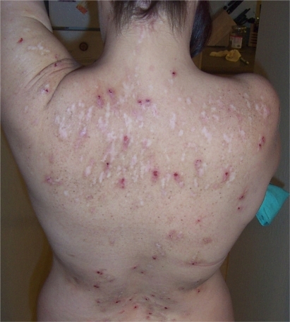
Figure 3 Close up of a leg lesion showing a twisted fiber just under the epidermis. Magnification 100 ×. Photo courtesy of Cindy Casey, Charles E Holman Foundation, Austin, Texas. Reproduced with permission.
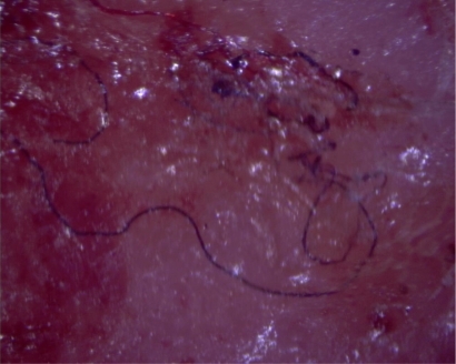
Figure 4 Black fibers and one red fiber, just under the epidermis of a healing lesion. Magnification 200 ×. Photo courtesy of Cindy Casey, Charles E Holman Foundation, Austin, Texas. Reproduced with permission.
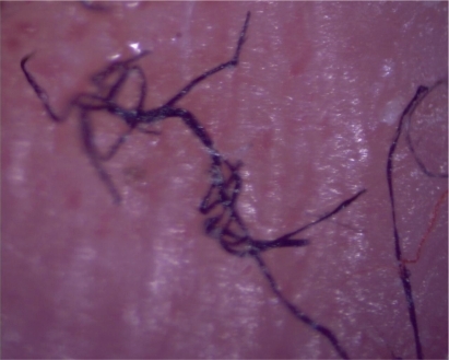
Figure 5 Fiber under epidermis (top) and separate from a skin lesion (bottom). Magnification 30 ×. Photo courtesy of Randy Wymore, Oklahoma State University Center for Health Sciences, Tulsa, Oklahoma. Reproduced with permission.
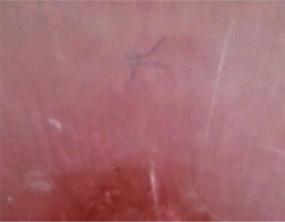
Table 1 Occupation of subjects at the time of presentation to the clinic (n = 122)
Table 2 Comorbidities of subjects (n = 122)
Table 3 Medications taken by subjects (n = 122)
Table 4 Events precipitating initiation of subjects’ symptoms (n = 122)
Table 5 Diagnoses given to study subjects prior to presentation to first author’s clinic (n = 122)
Table 6 Symptoms of patients with a positive exam for subcutaneous fibers (n = 122)
Table 7 Comparison of symptom prevalence in Morgellons group (n = 122) and Lyme group (n = 60)
Table 8 Chi square analysis of symptom prevalence in Morgellons group versus Lyme group