Figures & data
Figure 1 Lateral view of an orthognatic patient with Angle Class 2. The pictures show markedly different profiles. a) Correct position of the patient; b) tracings of photographs a, c, and d; c) the head is bent backward and the Frankfort Horizontal Plane is not parallel to the ground, and the deformity is therefore underestimated; d) the head is bent forward and the deformity is exaggerated.
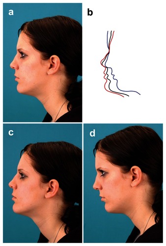
Figure 2 The 3dMD® cranial system uses 5 camera viewpoints to generate a 360° image of the head.
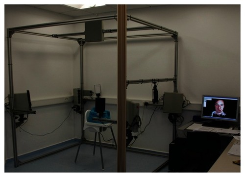
Figure 3 Five camera viewpoints of the head of a patient with deformational plagiocephaly. Camera views: a) half profile front right, b) half profile front left, c) half profile back left, d) half profile back right, e) from above.
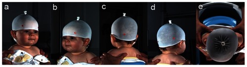
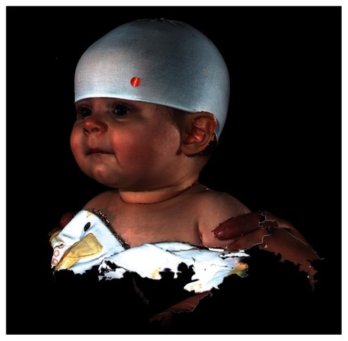
Figure 4 2D illustration of the composed 3D image of the patient’s head, which was generated from the 5 views in .