Figures & data
Figure 1 Spectrograms of the phrase “… raindrops in the air …” spoken by a 38 year old female: normal control subject is depicted on the top panel, ADSD before BT injection is on the middle, and ADSD after BT injection is on the bottom panel.
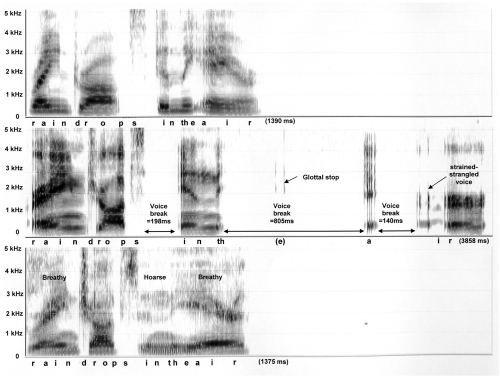
Figure 2 Spectrograms of the phrase “When the sunlight …” spoken by a 74 year old female: normal control subject is depicted on the top panel, ADSD before BT injection is on the middle, and ADSD after BT injection is on the bottom panel.
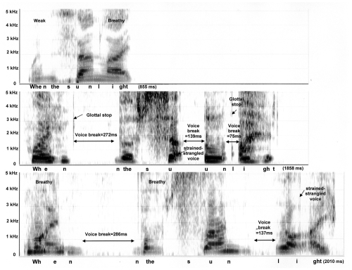
Figure 3 Coronal section of the larynx from 56 year old female illustrating major structural landmarks. Safran-Hematoxylin Stain, 2.5x original magnification. 1 – epiglottis, 2 – unossified thyroid cartilage, 3 – ossified portion of thyroid cartilage, 4 – unossified cricoid cartilage, 5 – ossified portion of cricoid cartilage, 6 – lamina propria of vocal fold, 7 – thyroarytenoid muscle (the major constrictor of vocal fold), 8 – laryngeal ventricle (lined of glandular epithelium), 9 – vestibular fold with G – mucous-serous glands, 10 – conus elasticus, 11 – glottal space.
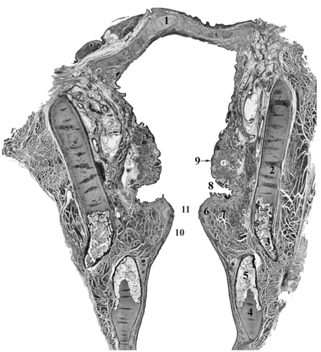
Figure 4 Coronal sections of the vocal fold from 21years old (A), 63 years old (B) and 72years old (C) females illustrating age related changes in the epithelium and lamina propria. Safran-Hematoxylin Stain, 20x original magnification. E – epithelium, S – superficial layer of the lamina propria, I – intermediate layer of the lamina propria, D – deep laryner of the lamina propria. Portions of the adjacent thyroarytenoid muscle (TA), which form a part of the body of the vocal fold, are also illustrated.
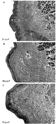
Figure 5 Endoscopic pictures of the vocal folds of a 58 year old woman during phonation, who presents with bowing of the vocal folds, thinning lamina propria and a persistent glottal chink.
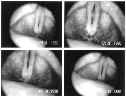
Figure 6 Diagrammatic representation of the major structures contributing to the peripheral speech mechanism. Structures of the upper and lower respiratory tracts are functionally interfaced and programmed by the CNS to produce voice and speech.
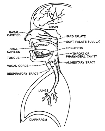
Table 1 Characteristics of speakers with adductor spasmodic dysphonia
Table 2 Attributes of voice and fluency for perceptual scaling experiments
Figure 7 Age-group plots of the means and standard deviations of visual analog scaling (VAS) scores for the four voice attributes (overall voice quality, roughness, brokenness, breathiness).
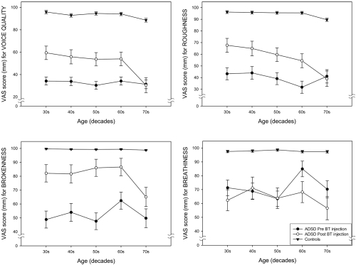
Figure 8 Age-group plots of the means and standard deviations of visual analog scaling (VAS) scores for the four fluency attributes (overall fluency, tension struggle, vocal spasms, dysfluent syllables).
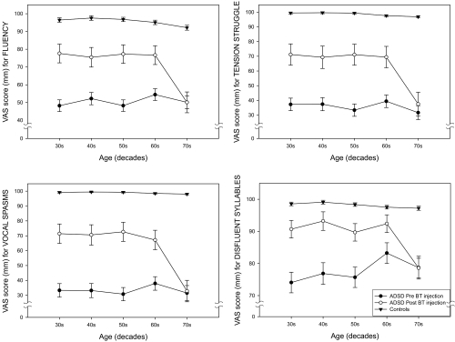
Table 3 Means and standard deviations of composite scores for voice, fluency, and overall scores are shown for three groups: controls, ADSD subjects before and after BT injection by age groupTable Footnote*
Figure 9 Interaction plots of decade by treatment condition for mean composite scores for voice for three groups: non-dysphonic controls, ADSD before BT injection and the same ADSD subjects after BT injection.
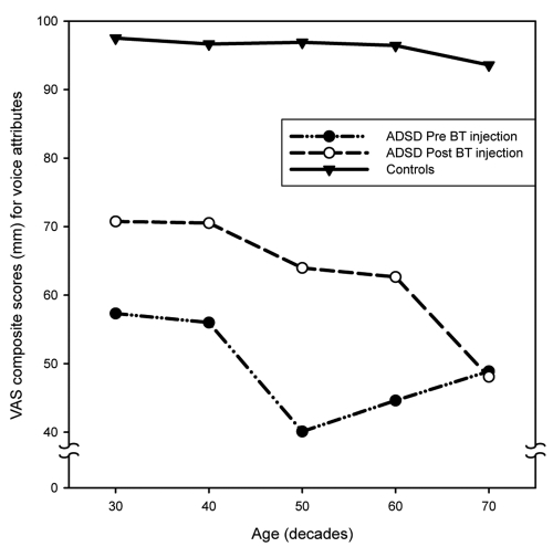
Figure 10 Interaction plots of decade by treatment condition for mean fluency composite scores by decade for three groups: non-dysphonic controls, ADSD before BT injection and the same ADSD subjects after BT injection.
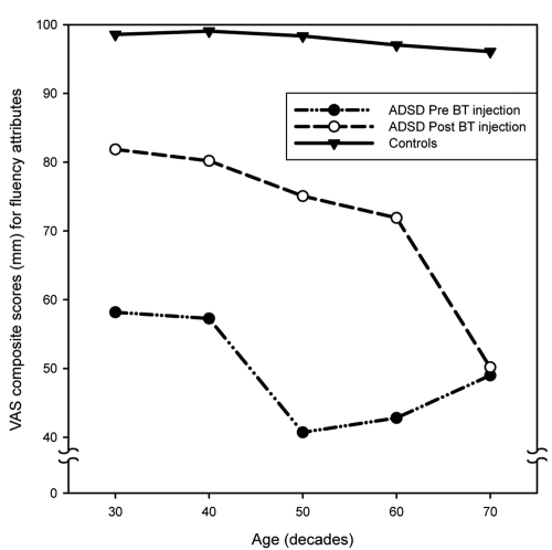
Table 4 Treatment characteristics for five age groups of ADSD