Figures & data
Figure 1 Low power view of biopsy specimen from lower lip from a 74-year-old woman who presented with a submucosal nodule of the lower lip. The patient had received injections of Restylane to the lips approximately 6 months before. Histologic examination of the biopsied specimen revealed the presence of multiple vacuolated, cyst-like areas. These are surrounded by a fibrotic connective tissue capsule and a tissue reaction composed predominantly of histiocytes and foamy macrophages. (Hematoxylin and eosin, original magnification 10×).
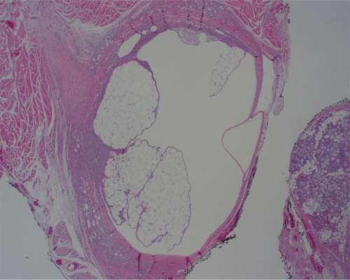
Figure 2 High power view of biopsy specimen from lower lip. Fibrotic tissue and abundant histiocytes surround the vacuolated areas. (Hematoxylin and eosin, original magnification 40×).
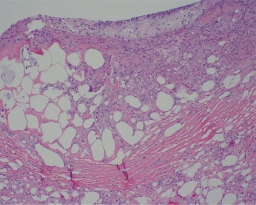
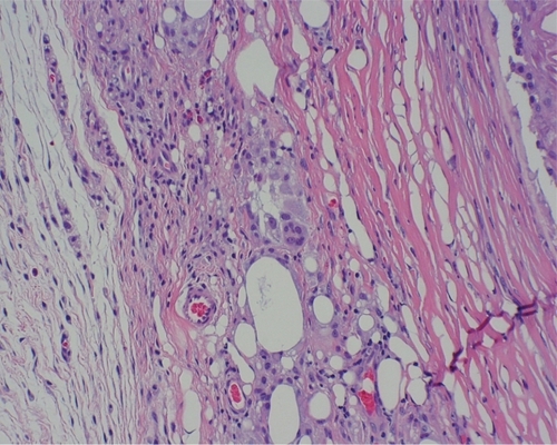
Figure 3 High power view of biopsy specimen from lower lip. A multinucleated foreign body-type giant cell is visible in the center of the photomicrograph. (Hematoxylin and eosin, original magnification 60×).