Figures & data
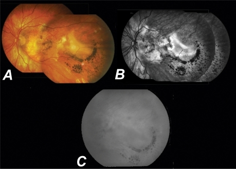
Figure 1 A) Color fundus picture of the right eye of our patient showing a macular scar. B) Color-mosaic fundus picture of the left eye (LE) before treatment showing a macular scar and a large hemorrhagic pigment epithelial detachment (PED) temporal to the fovea resembling a choroidal melanoma. C) Late phase of fluorescein angiography of the LE showing hyperfluorescence due to staining of fibrotic tissue at the fovea and hypofluorescence due to masking of the hemorrhagic PED temporal to the fovea. D) Mid phase of Indocyanine green angiography showing hypofluorescence due to the PED with two hot spots at the margins (white arrows).
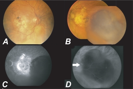
Figure 2 A) Color-mosaic fundus picture of the left eye (LE) six months post-ICGA-guided argon laser showing a macular scar and almost complete resolution of the hemorrhagic pigment epithelial detachment (PED) temporal to the fovea. The appearance remained stable for another six months. B) Late phase of FFA of the LE resolution of the PED temporal to the fovea with hyperfluorescence due to staining of the fibrotic tissue. C) Late phase of ICGA at the same time showing the area corresponding to the PED with no active hot spots.