Figures & data
Table 1 Age, serum CRP, MMP-3, VEGF levels before and after the treatment in patients with PMR, pseudogout, RS3PE syndrome, and post-infectious polyarthritis
Table 2 Changes in serum CRP levels after one week’s treatment using NSAIDs in patients with PMR, pseudogout, RS3PE syndrome, and post-infectious polyarthritis
Figure 1 Gallium-67 scintigraphic findings of patients with polymyalgia rheumatica (P), pseudogout A), remitting seronegative symmetrical synovitis with pitting edema (RS3PE) syndrome B), and post-infectious polyarthritis C). A–C corresponds to case A–C in , respectively. Arrows indicate a gallium uptake. Dotted arrows indicate a gallium uptake in metacarpophalangeal joints in a patient in RS3PE syndrome.
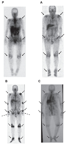
Figure 2 The radiographs of the patient with pseudogout demonstrated chondrocalcinosis (arrows) on the wrist A), knee B), and hip C).
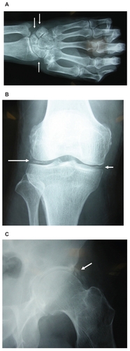
Table 3 Clinical and biochemical characteristics for polymyalgia rheumatic