Figures & data
Table 1 Studies of apoptosis in COPD
Table 2 Markers upregulating (↑) or downregulating (↓) apoptosis in COPD
Figure 1 Lung epithelial cell apoptosis. Increased apoptosis of epithelial cells may account for the decreased vascular endothelial growth factor (VEGF) expression, resulting in alveolar endothelial cells death. The compromised microcirculation resulting from endothelial cell damage may further promote pneumocyte cell death. Increased elastolytic activity in apoptotic lung epithelial cells amplifies the destruction of alveolar elastin. The subsequent destruction of the basement membrane and the loss of cell–extracellular matrix attachments lead to further apoptosis of the surrounding cells.
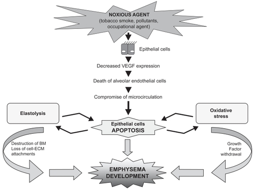
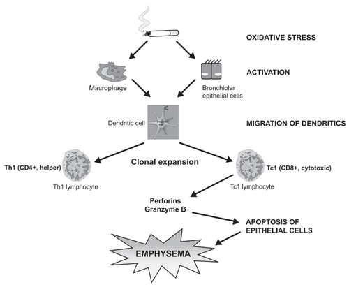
Figure 2 Oxidative stress of cigarette smoke in “susceptible” smokers activates adaptive immunity, predisposing dendritic cells to clonal expansion of CD4 + and especially of cytotoxic CD8 + cells, inducing epithelial cell death by perforin.