Figures & data
Table 1 Baseline characteristics
Figure 1 Mean basement membrane thickness for asthma, COPD, and healthy controls of similar age. Reticular basement membrane thickness in central airway wall biopsies from 24 patients with asthma, 12 age-matched healthy controls of asthma, 17 patients with COPD and 10 age- and pack year-matched healthy controls of COPD.
Abbreviation: COPD, chronic obstructive pulmonary disease.
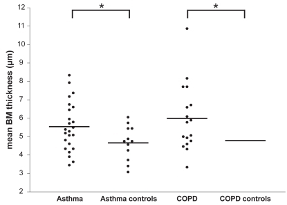
Figure 2 Reticular basement membrane composition. Relative staining density of collagen I, laminin, collagen IV, and tenascin in the reticular basement membrane of airway wall biopsies in asthma and COPD patients, and their healthy matched control subject. Results of collagen III and V are not shown as there were no significant differences between any of the groups.
Abbreviation: COPD, chronic obstructive pulmonary disease.
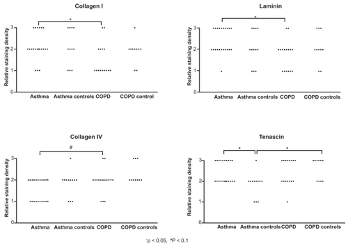
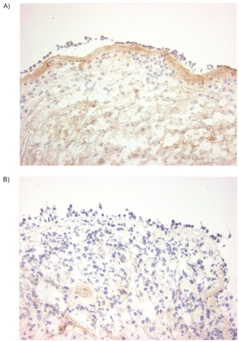
Figure 3 Collagen I staining. Immunohistochemical staining of bronchial biopsies for collagen I (immunoperoxidase, original magnification × 200) showed, a uniform red-brown-stained band beneath the epithelial layer in asthma (A left panel). In contrast to asthma, this is significantly less intense in COPD (p < 0.05, B right panel).