Figures & data
Figure 1 Representative light micrographs of sections (5 μm) of an uninjured (upper left panel) carotid artery from a rabbit inoculated with human arterial-derived nanoparticles, and the injured carotid artery of a rabbit inoculated with saline (control; upper right panel); calcified human arterial-derived (middle left panel) and calcified human kidney stone-derived (middle right panel and bottom right panel) nanoparticles; decalcified human arterial-derived nanoparticles (bottom left panel). All tissue was collected 5 weeks post-inoculation and sections are stained with either elastin van Giesen stain (upper and middle panels) or von Kossa stain (brown-black; lower panels). Sections are shown at the lowest (5X) magnification in order to show the entire artery. Uninjured arteries of rabbits in each treatment group are indistinguishable from that shown. Arrows indicate internal elastic lamina, bar indicates intimal thickening.
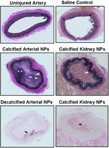
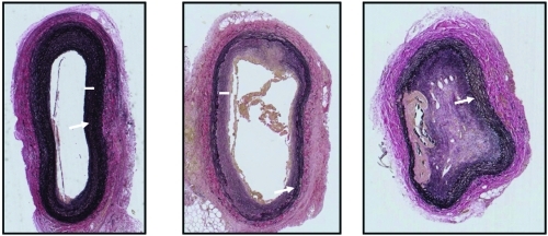
Figure 2 Representative light micrographs of sections (5 μm) of injured carotid arteries of three rabbits inoculated with decalcified human arterial-derived nanoparticles. All tissue was collected 5 weeks post-inoculation and stained with an elastin van Giesen stain. Sections are shown at the lowest (5X) magnification in order to show the entire artery. Uninjured arteries in these rabbits are indistinguishable from that shown in , upper left panel. Injured arteries from rabbits injected with decalcified human arterial-derived nanoparticles exhibited a spectrum of vascular healing, ranging from disruption of the internal elastic lamina with minimal development of intimal hyperplasia (left panel), disruption of the internal elastic lamina and greater intimal hyperplasia than developed in the saline-injected rabbits (middle panel), to complete occlusion with canalization and calcification (right panel). Arrows indicate internal elastic lamina, bars indicate intimal thickening.