Figures & data
Figure 1 CT abdomen: 11 × 10 × 6 cm cystic lesion with areas of rim calcification and few small cysts within it noted in the left lobe of liver extending into lesser sac. Mild ascites. Suggestive of hydatid disease of liver.
Abbreviation: CT, computed tomography.
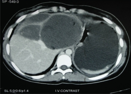
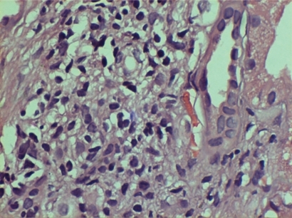
Figure 2 Glomeruli showed moderate mesangial matrix accentuation with hypercellularity. Capillary lumina were open with thickened, wrinkled membranes. Bowman’s capillary segmentally thickened and occasionally ruptured. Tubules had moderate degenerative changes. Mild interstitial edema with diffuse mixed leucocytic infiltration noted.
Note: Immunoflouresence showed nonspecific IgM trapping.
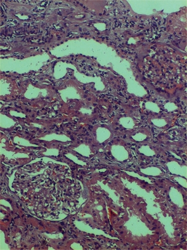
Figure 3 Acute on chronic tubulo-interstitial nephritis with mesangioproliferative glomerulonephritis.
Note: immunoflouresence showed nonspecific IgM trapping.