Figures & data
Figure 1 Images 7 months before the onset of pyogenic spondylodiscitis. A) Plain x-ray. Separation of the L4 vertebral arch (small white arrow) and L4/L5 listhesis were noted. B) Plain T2-weighted MRI. The L4/L5 intervertebral disc was degenerated and the height of the intervertebral disc space was decreased (thick white arrow). Separation of ring apophysis of the L4 vertebral body was noted (small black arrow).
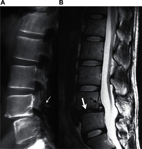
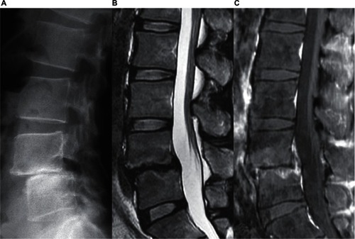
Figure 2 Images on admission. A) Plain x-ray. Although osteosclerosis of the L4 and L5 vertebral bodies was noted (small black arrow), no apparent bone destruction or spur formation was seen in the anterior corner of the vertebral bodies. B) Plain T2-weighted MRI. Signal intensity of the L4/L5 intervertebral disc had apparently changed in comparison with that shown in (thick white arrow). C) Gadolinium-enhanced fat-suppressed T1-weighted MRI. The L4 and L5 vertebral bodies adjacent to the L4/L5 intervertebral disc space were enhanced with gadolinium (small white arrow).
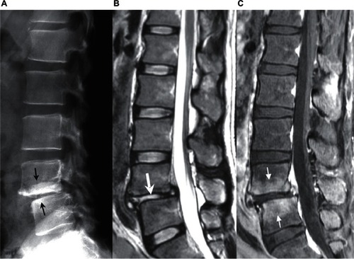
Table 1 Time courses of WBC count, neutrophil count, CRP, and ESR
Figure 3 Images 6 months after treatment. A) Plain x-ray. The L4/L5 intervertebral space was lost. B) Plain T2-weighted MRI. The high signal intensity of the L4/L5 intervertebral disc was resolved. C) Gadolinium-enhanced fat-suppressed T1-weighted MRI. Enhancement in the adjacent vertebral bodies remained.