Figures & data
Figure 1 Electrocardiogram revealing complete atrioventricular block with junctional escape rhythm (arrows).
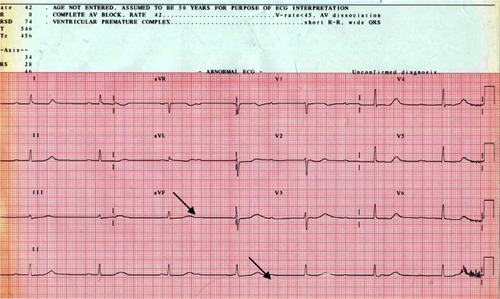
Figure 2 An ultrasonogram showing a massive infiltrative thickening of the posterior aspect of the left atrial septum and interatrial septum.
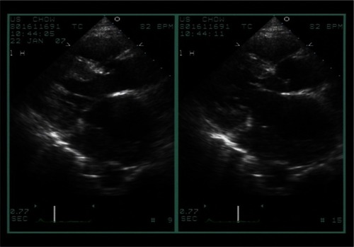
Figure 3 Transesophageal echocardiography showing tumor masses attached to the interatrial septum and protruding into both atrial cavities.
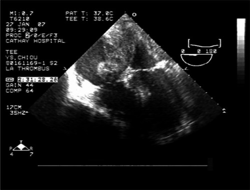
Figure 4 A multislice computed tomogram showing a bulky infiltrative mass about 8.62 cm × 6.45 cm × 9 cm at posterior-lateral and inferior aspect of the left atrium with invasion to myocardium, the interatrial septum, tricuspid valve, right atrium, pericardium, left hilar region, and left pleura (arrow).
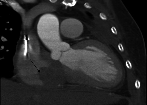
Figure 5 FDG-PET revealed remarkably increased glucose metabolism in the RA, interatrial septum, LA, and in a tumor mass protruding from the posterior aspect of pericardium.
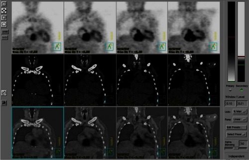
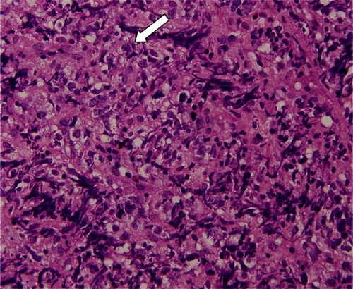
Figure 6 Section showing monotonous proliferation of large centroblast-like lymphoid cells having round to irregular nuclei, with vesicular chromatin and distinct 1 to 3 nucleoli and a moderate amount of cytoplasm (arrow).