Figures & data
Figure 1 Intracellular activation of ciclesonide and reversible esterification of des-CIC. Reprinted from CitationNave R, Meyer W, Fuhst R, et al 2005b. Formation of fatty acid conjugates of ciclesonide active metabolite in the rat lung after 4-week inhalation of ciclesonide. Pulm Pharmacol Ther, 18:390–6. Copyright © Elsevier.
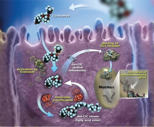
Table 1 Uptake of ciclesonide into A549 cells (derived from data of CitationNonaka et al 2007)
Table 2 Inhibition of esterases in HNEC and HNBE cells (derived from data of CitationMutch et al 2007; CitationSato et al 2007a)
Table 3 Formation of des-CIC fatty acid esters in airway epithelial cells and lung tissue
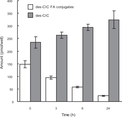
Figure 2 Reversible conjugation of des-CIC with fatty acid in human bronchial epithelial cells. Data were represented as the mean ± SD from 6 wells. Time point of 0 h indicates the end of incubation with des-CIC for 6 h. Results at 0 h represent only the amount in the cells and results at 3, 6, and 24 h represent the sum of analyte in the cells and medium harvested (Nonaka unpublished data).