Figures & data
Table 1 Characteristics of some lipid formulations under clinical trials.
Figure 1 Several pathways by which lipid formulations of AmB are thought to reach fungal or parasitic cells.
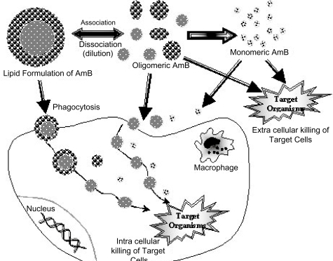
Figure 2 Several pathways by which the lipid formulations of AmB may reach mammalian cells.
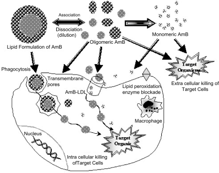
Figure 3 Proposed arrangement of AmB molecules (black) in the AmBisome bilayer. This structure accounts for the observation of rapid ion fluxes across the AmBisome bilayer in response to imposing a pH gradient from inside to outside. The individual AmB molecules form a “barrel” two of which fit tail-to-tail to form a pore spanning the bilayer. This structure is believed to contribute to the exceptional stability of AmBisome to loss of drug in buffer or plasma.
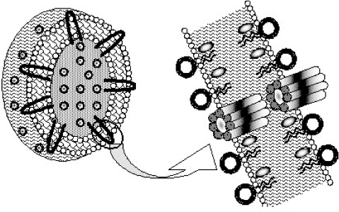
Figure 4 Antifungal activity of LNS-AmB, Fungizone, AmBisome, and DMSO-solubilized AmB in vitro. The growth inhibition of C. albicans was measured by the change in optical density at 540 nm in SD-MOPS broth after a 24-h incubation at 35°C. Results are the mean of two experiments.
Adapted from CitationFukui et al (2003).
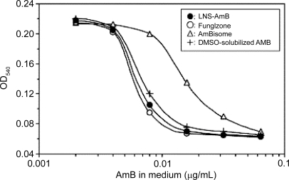
Figure 5 Survival of mice infected with C. albicans and treated with LNS-AmB, Fungizone, or AmBisome. Treatment was started 4 hours after fungal inoculation. +, P<0.05 compared with AmBisome; #, P<0.01 compared with Fungizone.
Adapted from CitationFukui et al (2003).
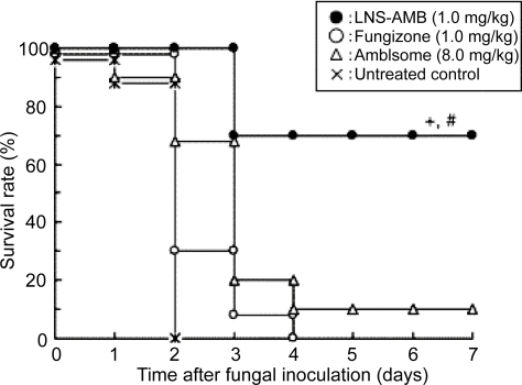
Figure 6 Photographs showing geimsa stained splenic smears of hamster treated with emulsomes and control formulations. A-untreated control group; B-Mycol (AmB for injection) treated group; C-TLEs orTrilaurin based emulsomes treated group; D-TSEs or Tristearin based emulsomes treated group.
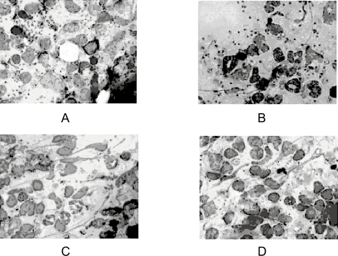
Table 2 Activity of emulsome formulations against L. donovani in hamsters infected for 30 days