Figures & data
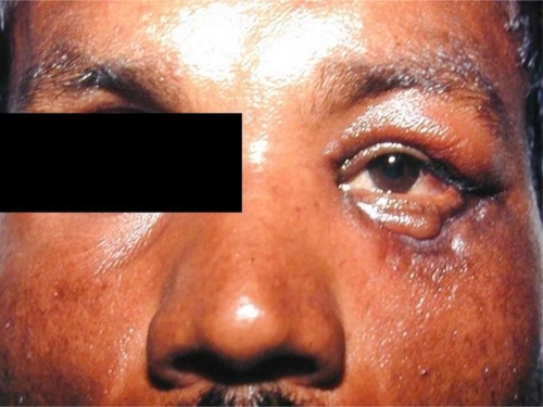
Figure 1 Brawny nonpitting edema of the upper and lower eyelids of the left eye with serosanguinous discharge
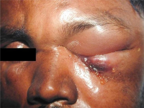
Figure 2 Gel electrophoresis for B. anthracis.
Notes: Lane 1 and 9- PA and CAP (positive control); Lane 2 and 10- PA and CAP (negative control); Lane 3 and 11- undiluted DNA for PA and CAP (serous fluid); Lane 4 and 12- 1/10 diluted DNA for PA and CAP (serous fluid); Lane 5 and 13- water blank; Lane 6 and 14- undiluted DNA for PA and CAP (blood); Lane 7 and 15- 1/10 diluted DNA for PA and CAP; Lane 8- marker (100-bp ladder).
Figure 3 Black eschar involving the left lower lid with residual edema of the left upper lid.
Figure 4 Ulcerated undersurface of the left lower lid on removal of the eschar.

Figure 5 Scarring and ectropion of the left lower lid.