Figures & data
Figure 1 Fundus photography with a line showing the OCT scan. The photograph demonstrates the presence of a tilted disc surrounded by a slightly elevated yellow-orange lesion, consistent with a PPRD, which hampered visualization of the underlying choroidal vessels.
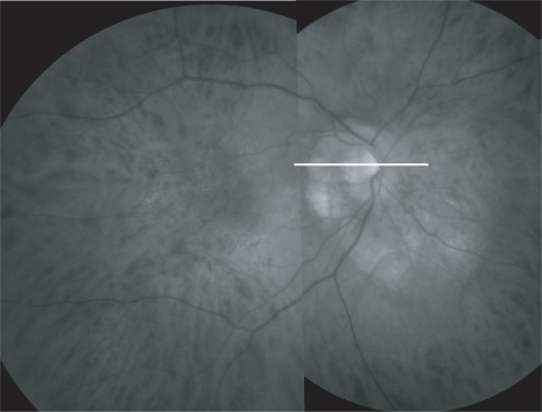
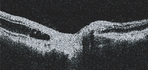
Figure 2 The OCT image of the right eye demonstrates the presence of a nonreflective space between the RPE and the neurosensory retina all around the optic disc. The presence of some bridging tissue (probably Muller’s cells) led us to classify it as an outer retinal layer schisis. No epiretinal membranes were detected. The scans crossing the optic nerve head confirmed the lack of an optic disc pit.