Figures & data
Table 1 Patient characteristics and results of combined photodynamic therapy and intravitreal bevacizumab
Figure 1 (a) and (a-1) Pretreatment fundus photo and pretreatment FLA, late phase, showing filling of predominantly classic CNV occupying more than 50% of the lesion. Pretreatment BCVA = 20/400. (b) and (b-1) Post-treatment fundus photo and FLA, late phase, at 6 months showing closure of the CNV with only late leakage at its upper temporal edge (BCVA = 20/150). (c) Pretreatment OCT showing CNV with CRT = 558 μm. (c-1) OCT, 6 months post-treatment showing absorption of most of subretinal fluid and reduction of CRT to 248 μm. However, fovea did not return completely to normal configuration.
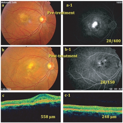
Figure 2 (a) Pretreatment fundus photo showing juxtafoveal CNV with subretinal hemorrhage occupying about 50% of the lesion (BCVA = 20/400). (a-1) Post-treatment fundus photo at 6 months. Note regression of the membrane and absorption of most subretinal blood (BCVA = 20/40). (b) Pretreatment FLA, showing active CNV. (b-1) Post-treatment FLA showing CNV staining with no leakage. (c) Pretreatment OCT showing the CNV with fluid in the subretinal space (CRT = 800 μm). (d) OCT 6 months post-treatment with almost normal foveal configuration (CRT = 280 μm).
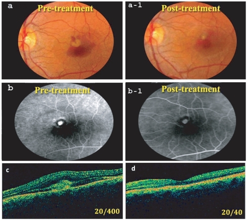
Table 2 Choroidal neovascularization subtype characteristics