Figures & data
Figure 1 Initial right eye slit-lamp examination. The nasal bulbar and inferior conjunctivas (A) and the superior tarsal conjunctiva (B) had plaque-like ulcerations. The initial conjunctival ulceration was followed by a recurrence of the inferior tarsal ulcer (C). A nasal symblepharon remained as a sequela after a 9-month followup (D).
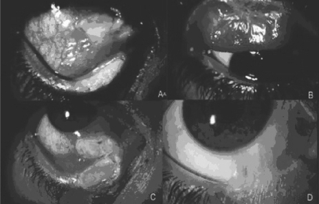
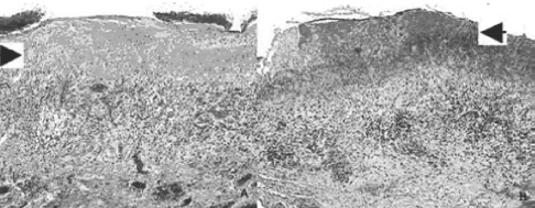
Figure 2 Histopathology of conjunctival biopsies. The superficial ulceration had an underlying thick band of hyaline-like eosinophilic material, probably composed of fibrin (arrow). Granulation tissue and lymphocytic infiltration were apparent in H&E (A) and fibrin (arrow) was confirmed with Lendrum staining (B). The tissue sample was negative for Congo red.