Figures & data
Figure 1 Light photomicrographs of histological section obtained from the periphery of the corneal button (hematoxylin and eosin stain). Photomicrograph showing detached epithelial layer and attenuated endothelial layer. Arrows indicate the interfaced between the host and donor stroma. Original magnification × 40.
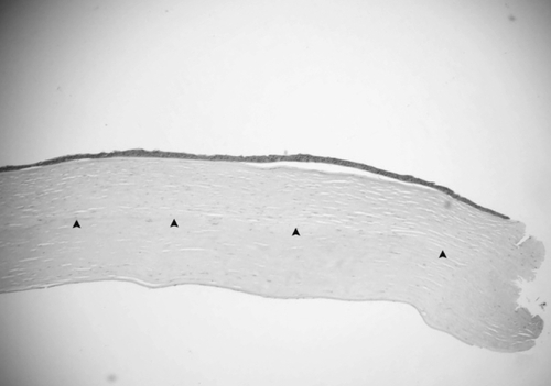
Figure 2 Light photomicrographs of histological section obtained from the center of the corneal button (hematoxylin and eosin stain). Photomicrograph showing attached corneal stoma. Original magnification × 40.
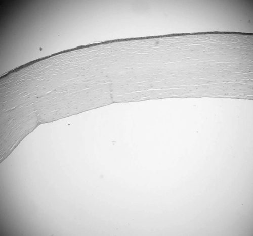
Figure 3 Light photomicrographs of histological section obtained from the periphery of the corneal button (hematoxylin and eosin stain). Photomicrograph showing stroma separation with a piece of Descemet’s membrane (arrows) and an amorphous fragment (stars). Original magnification × 100.
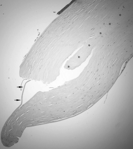
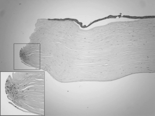
Figure 4 Light photomicrographs of histological section obtained from the periphery of the corneal button (hematoxylin and eosin stain). Photomicrograph showing increased keratocytes at the posterior edge. Original magnification × 100. Inserted figure manifested high magnification × 200.