Figures & data
Table 1 Age, follow-up, and distribution of cases according to surgical diagnosis
Figure 1 Fundus photographs of a case in group 2. A before injection of bevacizumab showing vitreous and subhyaloid hemorrhage and tractional retinal detachment. B 1 week after injection of bevacizumab showing evident regression of retinal neovascularization and C 12 months after vitrectomy with retina attached with near confluent laser marks.
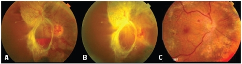
Table 2 Intraoperative and postoperative data