Figures & data
Table 1 Clinical characteristics at baseline
Figure 1 The stent is seen correctly placed at the trabecular meshwork level.
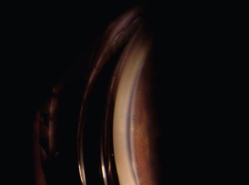
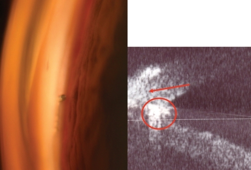
Figure 2 Left: The gonioscopic view of the stent. Right: The UBM shows the hyperechogenic structure of the stent (circle), placed below the corneal wedge (arrow).