Figures & data
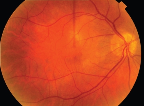
Figure 1 Fundus appearance of the right eye at initial presentation. There was a larger yellowish placoid lesion, with a small splinter haemorrhage in the macular region and mild vitreous inflammation.
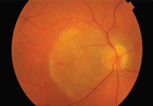
Figure 2 A: Fundus fluorescein angiogram demonstrating early hypofluorescence in the affected area. B: There was late staining with diffuse, non-progressive hyperfluorescence. There were no signs of a choroidal neovascular membrane.
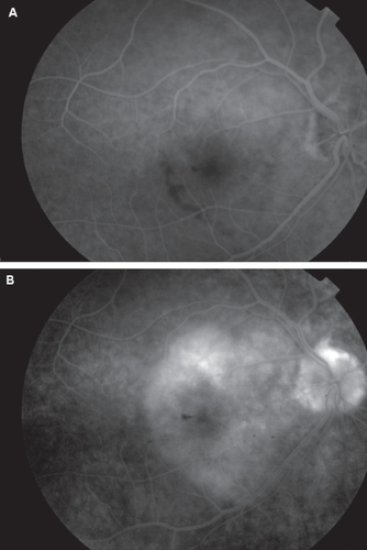
Figure 3 An OCT scan of the right eye at initial presentation. There was some thickening of the RPE layer but there were no signs of any retinal oedema or serous detachment.
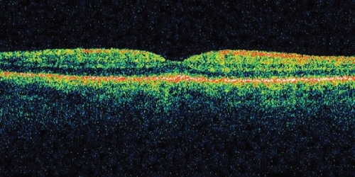
Figure 4 Fundus appearance of the unaffected left eye.
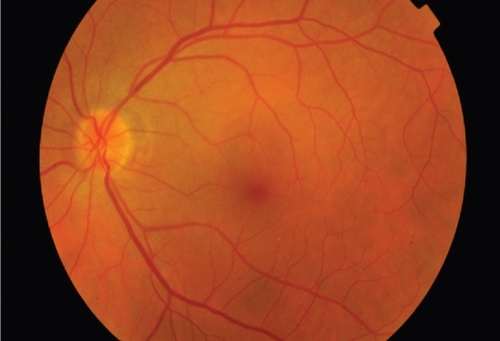
Figure 5 Fundus appearance at 5-month follow-up. The retina had returned to normal apart from underlying chorioretinal atrophy in the region of the previous placoid infection.