Figures & data
Figure 1 The preoperative visual field. A) Left eye. B) Right eye.
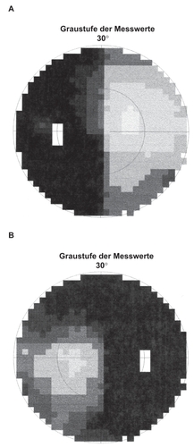
Figure 2 MRI, coronal section, T2-weighted image: the enlargement of the optical chiasm can easily be seen.
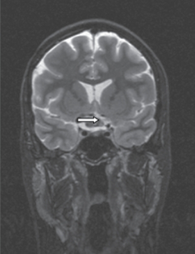
Figure 3 A) MRI, axial section, T1-weighted images after administration of gadolinium: the irregular contrast enhancement (arrow) can be seen. B) MRI, sagittal section, T1-weighted image after administration of gadolinium: the enlarged chiasma suprasellar above the pituitary gland can be seen.
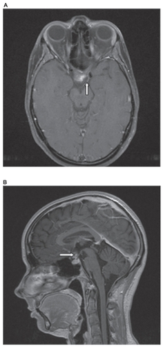
Figure 4 A) Intraoperative photograph after pterional approach from the right-hand side and splitting of the sylvian fissure: blue scarring in the region of lamina terminalis can be seen together with a bulging and thinning of the right-hand optic nerve (arrow). B) Intraoperative photograph after pterional approach from the right-hand side: the focused contralateral yellow optic nerve (arrow) seems uninvolved by the lesion with normal vascularization.
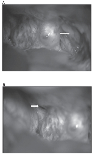
Figure 5 Intraoperative photograph after pterional approach from the right-hand side and resection of the cavernoma. The suction tube touches the beginning of the left optic nerve. One can look through the right-hand optic nerve, because it is extremely thin.
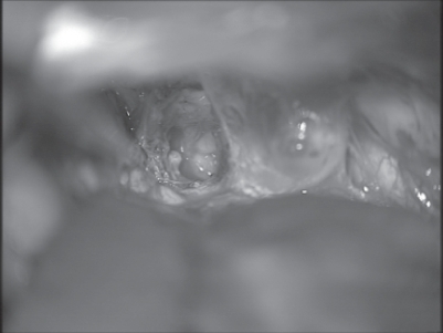
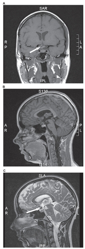
Figure 6 Postoperative MRI. A) MRI, coronal section, T1-weighted images: a postoperative defect inside the chiasm is visible. B) MRI, sagittal section, T1-weighted images after administration of gadolinium: no residual cavernoma is visible. C) MRI, sagittal section, T2-weighted images revealing complete resection of the cavernoma with minimal hemosiderin residuals.