Figures & data
Figure 1 Time course of the number of Fluoro-Jade B-positive cells in the GCL. The number of Fluoro-Jade B positive cells in the GCL peaks at 2 days after reperfusion.
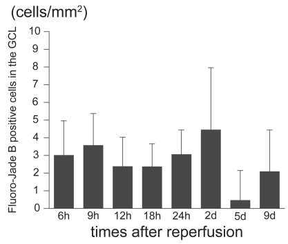
Figure 2 Expression of IRE1α in control and ischemic retinas. The section was obtained from the ischemic retina 6 hours after reperfusion. In the ischemic retina, the expression of IRE1α in the GCL is significantly stronger than in the control retinas (A) (merged). Although part of the IRE1α expression was detected with Fluoro-Jade B positive signals (B) (multi panel), most IRE1α expression did not co-exist with Fluoro-Jade B positive signals (A).
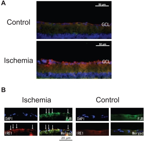
Figure 3 Intensities of IRE1α expression in control and ischemic retinas. Texas-Red intensities of IRE1α expression in the GCL of ischemic retinas are significantly increased compared to that of the control retinas (179.8 ± 16.3 pixels/mm2 versus 147.6 ± 26.9 pixels/mm2, P = 0.0011 by Mann Whitney U test).
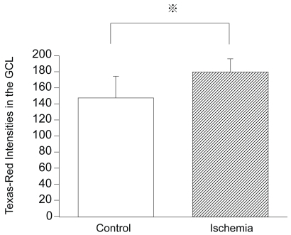
Figure 4 Western blot analysis of IRE1α protein levels in ischemic retinas. In the ischemic retinas, the IRE1α protein level was significantly increased compared to that of the control retinas (100 ± 15.9% of control versus 159.9 ± 73.9% of control, P = 0.0369 by Mann-Whitney U test). All densitometric values for western blot analysis were normalized with β-actin signals.
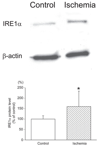
Figure 5 ASK1 expression co-localized with Fluoro-Jade B-positive cells in ischemic retinas and the time course of the number of ASK1-positive cells that are also Fluoro-Jade B-positive. The number of ASK1-positive cells and the number of ASK1 positive cells co-colocalized with Fluoro-Jade B positive signals in the ischemic retinas were significantly increased compared to that of the control retinas only after 6 hours of reperfusion (A). In ischemic retinas, more than one-half of the cells that were ASK1-positive were also Fluoro-Jade B-positive in the GCL (B). The sections were obtained from an ischemic retina 6 hour after reperfusion (C) (multi panel). The expression of ASK1 co-existed with Fluoro-Jade B-positive cells in the GCL (arrows) (C). (A); White bars showed the number of ASK1 positive cells in the control retinas and checked bars showed the number of ASK1 positive and Fluoro-Jade B positive cells in the control retinas. Black bars showed the number of ASK1 positive cells in the ischemic retinas and stripe bars showed ASK1 positive and Fluoro-Jade B positive cells in the ischemic retinas. C, control; I, Ischemia; C/F, Control (ASK1) co-existed with Fluoro-Jade B; I/F, Ischemia (ASK1) co-existed with Fluoro-Jade B.
Abbvreviations: h, hours; d, days.
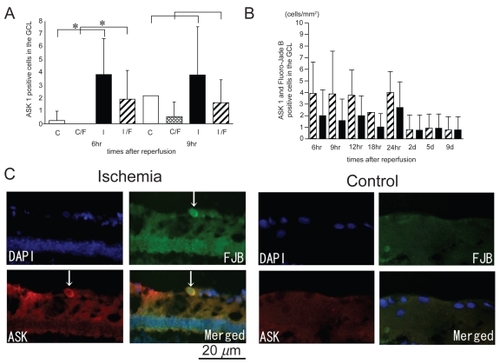
Figure 6 P-SEK1/MEK4 expression co-localized with Fluoro-Jade B-positive cells in the GCL and the time course of the number of p-SEK1/MEK4 positive cells in the GCL. The numbers of p-SEK1/MEK4 positive cells in the GCL in the ischemic retinas were significantly increased compared to those of the control retinas only after 6 hours and 9 hours of reperfusion (A). The number of p-SEK1/MEK4 positive and Fluoro-Jade B positive cells in the GCL in the ischemic retinas was significantly increased compared to that of the control only after 6 hours of reperfusion (A). In ischemic retinas, more than one-half of the cells that were p-SEK1/MEK4-positive were also Fluoro-Jade B-positive in the GCL (B). The sections were obtained from ischemic retinas 6 hours after reperfusion (C) (multi panel). The expression of p-SEK1/MEK4 is co-localized with Fluoro-Jade B-positive cells in the GCL (arrows) (C). (A); White bars showed the number of p-SEK1/MEK4 positive cells in the control retinas and checked bars showed the number of p-SEK1/MEK4 positive and Fluoro-Jade B positive cells in the control retinas. Black bars showed the number of p-SEK1/MEK4 positive cells in the ischemic retinas and stripe bars showed p-SEK1/MEK4 positive and Fluoro-Jade B positive cells in the ischemic retinas.
Notes: *P < 0.05 by Mann Whitney U test. (B); Stripe bars showed the number of p-SEK1/MEK4 positive cells and black bars showed the number of p-SEK1/MEK4 positive cells co-existed with Fluoro-Jade B-positive cells in the GCL of ischemic retinas.
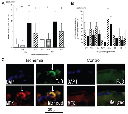
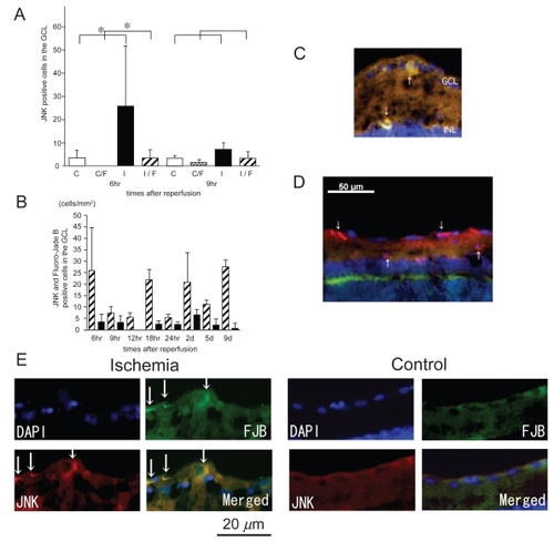
Figure 7 Expression of p-JNK in ischemic retinas and the time course of the number of p-JNK positive cells in the GCL. The numbers of p-JNK positive cells and p-JNK/Fluoro-Jade B positive cells in the ischemic retinas were significantly increased compared to those of the control retinas only after 6 hours of reperfusion (A). Less than one-half of the p-JNK-positive cells were also Fluoro-Jade B-positive (B). The sections were obtained from ischemic retinas 6 hours after reperfusion (C–E). The expression of p-JNK co-existed with Fluoro-Jade B-positive cells in the GCL and the INL (arrows) (C) (merged) and (E) (multi panel). However, many type of cells with no Fluoro-Jade B positive signals were p-JNK-positive (D) (merged). Thus, JNK may play different roles in ischemic retinas as well as neuronal degeneration. (A); White bars showed the number of p-JNK positive cells in the control retinas and checked bars showed the number of p-JNK positive and Fluoro-Jade B positive cells in the control retinas. Black bars showed the number of p-JNK positive cells in the ischemic retinas and stripe bars showed p-JNK positive and Fluoro-Jade B positive cells in the ischemic retinas.
Notes: *P < 0.05 by Mann Whitney U test. (B); Stripe bars showed the number of p-JNK positive cells and black bars showed the number of p-JNK positive cells co-localized with Fluoro-Jade B-positive cells in the GCL of ischemic retinas.