Figures & data
Figure 1 This shows the degree of extraocular muscle thickening in coronal slice pre- and poststeroid pulse treatment.
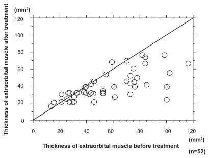
Figure 2 We gathered serum from pre- and poststeroid pulse treatment patients and measured sFas/IL-6. A shows the changes in sFas value. Student t-test revealed the value of sFas to be significantly decreased following steroid pulse treatment (student t test, p < 0.01). B shows the changes in IL-6. IL-6 did not decrease in either pre- or poststeroid pulse treatment.
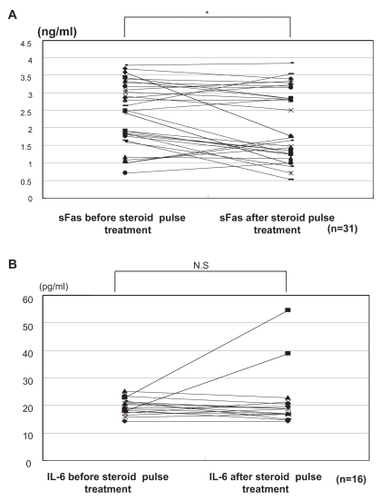
Figure 3 This shows the serum level of sFas before and after steroid pulse treatment.
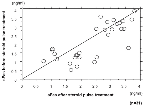
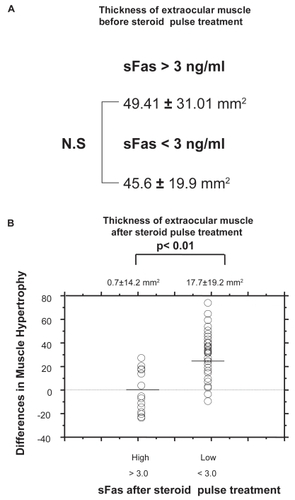
Figure 4 A shows the cross-section area of extraocular muscle before steroid pulse treatment. No significant difference in the cross-section area of extraocular muscle was observed between subjects with sFas levels of less than 3 ng/ml and those with more than 3 ng/ml. B shows those with sFas level of less than 3 ng/ml after steroid pulse treatment disclosed improvement in extraocular muscle thickness (Man-Whitney’s U test).