Figures & data
Figure 1 (A) The effect of contact lens wear on basal cell proliferation. Single pulse BrdU labeling in the rabbit corneal epithelium following 24 hours of contact lens wear demonstrated a reduction in basal cells mitosis in all lens groups tested and from hypoxia induced by eyelid suturing, suggestive of both lens- and oxygen-mediated effects. (B) The effect of contact lens wear on apoptosis in the central corneal epithelium. Annexin V labeling, an early marker for apoptosis, also showed a reduction in epithelial desquamation with all forms of contact lens wear, which did not appear to be related to lens-oxygen transmissibility. Copyright © 2008. Figures adapted with permission from CitationLadage PM, Yamamoto K, Li L, et al. 2002. Corneal epithelial homeostasis following daily and overnight lens wear. Contact Lens Anterior Eye, 25:11–21.
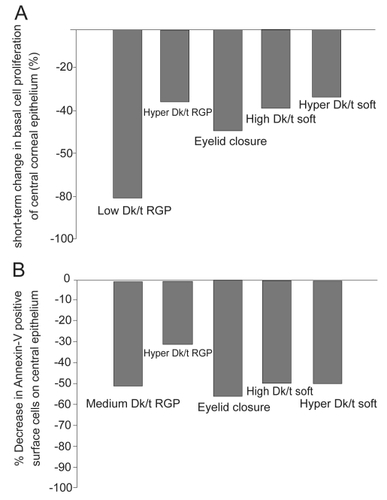
Figure 2 (A) Percentage of epithelial desquamation as a function of equivalent oxygen percentage (EOP). As lens oxygen transmissibility increased, there was a significant decrease in epithelial desquamation for both RGP and hydrogel lenses. At lower oxygen transmission levels, RGP lenses induced greater levels of surface epithelial desquamation. At the highest EOP tested, RGP lenses induced no observable desquamation. (B) Increase in PA adherence, expressed as colony forming units (CFU), as a function of EOP. Similar to desquamation, increasing EOP levels resulted in a reduction of PA adherence. In contrast to desquamation, for each level of oxygen transmission, rigid lenses bound significantly less PA than hydrogel lenses. Copyright © 1994. Figures adapted with permission from CitationImayasu M, Petroll WM, Jester JV, et al. 1994. The relation between contact lens oxygen transmissibility and binding of Pseudomonas aeruginosa to the cornea after overnight wear. Ophthalmology, 101:371–88.
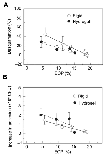
Figure 3 Lipid raft expression in the rabbit corneal epithelium. After 24 hours of PMMA lens wear, whole mount tissue was labeled with beta cholera toxin (green) and counterstained with PI (red) to label epithelial nuclei and allow for visualization of PA. (A) Undisturbed corneal epithelium. No lipid raft expression was seen in the absence of a contact lens. (B) After 24 hours of lens wear, pre-infection with PA, lipid rafts were detected in surface epithelial cells, evident by green punctate staining. (C)Thirty minutes after infection with PA, PA were seen to preferentially adhere to lipid raft expressing cells. (D) At 1 hour post infection, PA appeared to cluster around the lipid raft fraction of the plasma membrane. In the vertical XZ plane, lipid rafts appeared to associate directly with the lipid raft and internalization was noted. Copyright © 2005. Image adapted with permission from CitationYamamoto N, Yamamoto N, Petroll WM, et al. 2005. Internalization of Pseudomonas aeruginosa is mediated by lipid rafts in contact lens-wearing rabbit and cultured human corneal epithelial cells. Invest Ophthalmol Vis Sci, 46:1348–55.
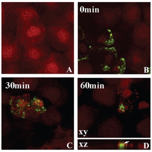
Figure 4 (A) Schematic of the noninvasive ocular irrigation device. Warmed saline is gently applied to the eye and collected in a test tube directly below. Cells are then incubated in an invasive strain of PA for 30 minutes, vacuum filtered onto polycarbonate filters, and stained with acridine orange for visualization using an epifluorescent microscope. (B) Correlation of PA adherence between exfoliated corneal epithelial cells and the residual corneal surface (R = 0.78, P < 0.001). Copyright © 1997. Images adapted with permission from CitationRen DH, Petroll WM, Jester JV, et al. 1997. Adherence of Pseudomonas aeruginosa to shed rabbit corneal epithelial cells after overnight wear of contact lenses. CLAO J, 23:63–8.
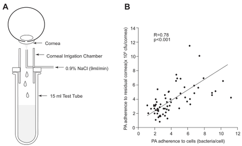
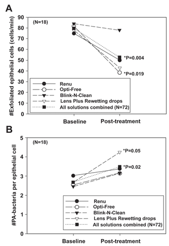
Figure 5 (A) The number of exfoliated cells (cells/min) before and after treatment. Surface cell exfoliation was reduced after repeated exposure to chemically preserved MPS. In addition, Lens Plus which contains the buffering agent boric acid, also showed a significant effect on the decrease in the number of cells shed from the epithelial surface. (B) The number of PA bound per epithelial cell before and after treatment. Concurrent with a decrease in desquamation, there was an increase in PA adherence seen with all solutions tested. Copyright © 2003. Figures adapted with permission from CitationLi SL, Ladage PM, Yamamoto T, et al. 2003. Effects of contact lens care solutions on surface exfoliation and bacterial binding to corneal epithelial cells. Eye Contact Lens, 29:27–30.