Figures & data
Figure 1 (A) Percent hemolysis (n = 3 per strain) for study strains. Percent was determined by comparison to a saponin 100% control. None of the high hemolytic strains (1, 2, 3) were significantly different from each other (P > 0.05). All of the high hemolytic strains were significantly higher than the low hemolytic strains (4 and 5) (P < 0.05). Strain 4 was significantly lower than strain 5 (P < 0.0001) Error bars denote standard deviation. (B) Concentration of PLY for study strains as determined by ELISA. Error bars denote standard deviation. Strain 3 had significantly more PLY than all other strains (P < 0.05). Strains 1 and 5 had significantly more PLY than strains 2 and 4 (P < 0.05). Strains 1 and 5 and strains 2 and 4 are not significantly different (P > 0.05).
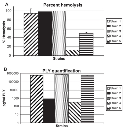
Figure 2 Average SLE scores at 24 hours PI. All strains producing high hemolytic activity in vitro (strains 1, 2, and 3) caused significantly higher SLE scores in vivo than the low activity strains (strains 4 and 5; P < 0.05). There was no significant difference in SLE scores between the two low activity strains (P = 0.8342). There were no significant differences in SLE scores between the three high activity strains (P > 0.05), except for strain 1 compared to strain 3 (P = 0.0352). Error bars denote standard errors of the means.
Abbrevations: PI, post infection; SLE, slit lamp examination.
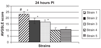
Figure 3 Pictures of representative eyes at 24 hours PI (A) and 48 hours PI (B). Strains 3 (left column) and 5 (right column) are shown. N = 12 for all strains except strain 4 (n = 10).
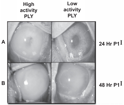
Table 1 Log10 CFU recovered from the vitreous
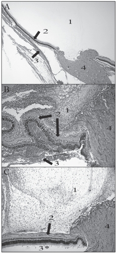
Figure 4 Representative histology pictures of an uninfected eye (A; 20X magnification) and eyes infected with strains producing high (B; 40X magnification) or low (C; 40X magnification) PLY activity. 1 is vitreous; 2 is retinal layers; 3 is choroid; 4 is optic disk. (A) Normal vitreous (1), retina (top arrow), and choroid (bottom arrow) were observed in an uninfected eye. (B) Damage and inflammation of the retina were observed (top two arrows). The choroid had become separated from the retinal layers by PMNs (bottom arrow). Severe inflammation was observed in the vitreous (1). (C) Only moderate inflammation was observed in the vitreous (1). Minor inflammation was observed in the retinal layers (arrow).
Abbreviations: PLY, pneumolysin; PMNs, polymorphonuclear cells.