Figures & data
Table 1 Case distribution
Table 2 Genetically determined cases
Table 3 Age of onset
Figure 1 Retinal photograph OS showing widespread pigmentary clumping (boney spicule changes) in the peripheral fields and attenuation of the vessels. OD was perfectly normal.
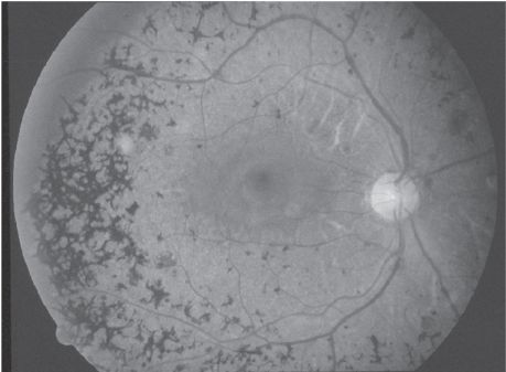
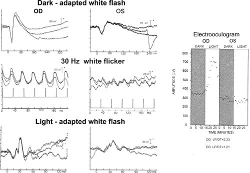
Figure 2 Electroretinogram shows normal responses OD and an absent response OS to blue flash. White flash in both the dark-adapted and light-adapted states have marked reduced a- and b-waves with prolonged implicit times. This pattern is consistent with moderately advanced retinitis pigmentosa with the rod responses being more affected than the cones. The electrooculogram shows a normal light response OD and no response to light OS. These results confirm severe damage to the retinal pigment epithelial layer.
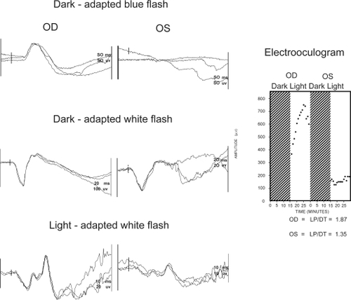
Figure 3 Electroretinogram and electrooculogram in a more advanced case of unilateral retinitis pigmentosa. In this example the affected eye is OD, where no responses to any stimuli elicited. OS has normal responses in both the dark-adapted and light-adapted states. The electrooculogram demonstrates no response to light OD while OS has a normal response to light.
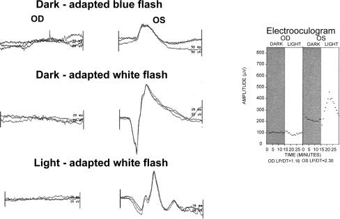
Figure 4 Electroretinogram and electrooculogram in one of the individuals with unilateral cone-rod dystrophy. OS is the affected eye and shows residual rod function to white flash in the dark-adapted state. The a- and b-waves are reduced in amplitude and the implicit times prolonged. The b-wave is poorly formed, reduced in amplitude and has a prolonged implicit time to 30 Hz white flicker. White flash in the light-adapted state shows abnormally small a- and b-waves with prolonged implicit times. The electrooculogram demonstrates no response to light OS and a normal response OD.