Figures & data
Figure 1 Rundle’s curve mapping increase in disease activity or severity followed by a reduction over time.
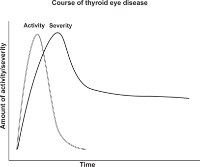
Figure 2 Severe inflammation and proptosis with classic “stare” of thyroid eye disease may be prominent in the active phase of disease.
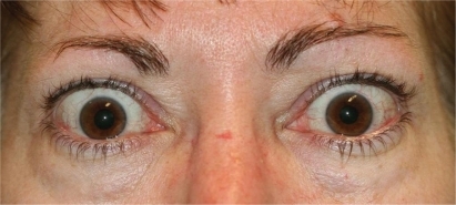
Figure 3 Esotropia strabismus is a common manifestation of medial rectus enlargement in thyroid eye disease.
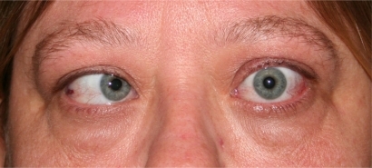
Figure 4 CT imaging of extraocular muscle enlargement at orbital apex.
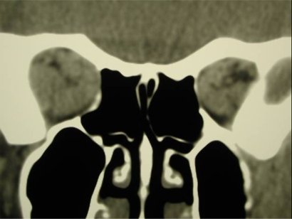
Table 1 Clinical activity or severity may be assessed with either the Clinical Activity Score (CAS) or the NOSPECS severity assessment
Table 2 Proposed methyprednisolone dosing regimens
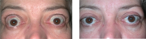
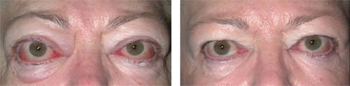
Figures 5 (left) and 6 (right) Pre- and post-operative orbital decompression images document a marked decrease in proptosis.
Figures 7 (left) and 8 (right) Pre- and post-operative strabismus surgery images document improvement of motor alignment.
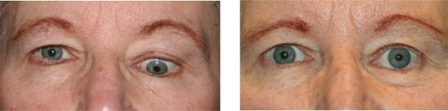
Figures 9 (left) and 10 (right) Pre- and post-operative eyelid retraction repair images document improvement of eyelid position.