Figures & data
Figure 1 Computed tomographic images showing blow-out fracture of the lower right orbital wall. Left: Coronal section showing that the right eyeball is not perforated but has shifted downward into the maxillary sinus through the fracture of the lower orbital wall. right: Axial section showing that her right eyeball was displaced.
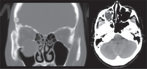
Figure 2 Postoperative computed tomographic image and fundus photograph. Left: Postoperative computed tomographic image of axial section. Blow-out fracture has been corrected and the eyeball is returned to the proper position. right: Fundus photograph of right eye showing a cherry-red spot, milky-white lesion on macula, and mild venous dilation.
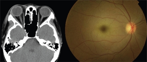
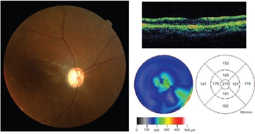
Figure 3 Optical coherence tomographic image and fundus photograph one month after the accident. Left: Fundus photograph of right eye showing optic atrophy. right: Axial slice (upper) and retinal thickness analysis (lower) of optical coherence tomographic image.