Figures & data
Figure 1 Diagrammatic representation of the modified retrograde dacryocystorhinostomy technique. A blunt-tipped ‘O’ gauge probe, the distal 1.5 cm of which is at right angle, is inserted retrogradely into the common internal opening and passed in the direction of the lower canalicular system. Note the scalpel which is used to cut down onto the probe tip and create a pseudopunctum.
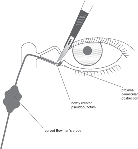
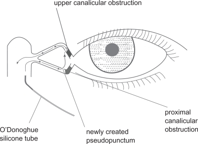
Figure 2 Diagrammatic representation of the modified retrograde dacryocystorhinostomy technique. O’Donoghue silicone intubation is performed, the stent being introduced through the common ostium, out through the pseudopuncum of the lower canaliculus and returned through the punctum of the normal upper canaliculus down through the common ostium into the nose.

Figure 3 Diagrammatic representation of the modified retrograde dacryocystorhinostomy technique. In cases where both canaliculi are obstructed, we advocate introducing the stent through the common internal opening into either canaliculus (herein lower canaliculus) and then out from one pseudopunctum into the other before returning into the nose.