Figures & data
Figure 1 Ophthalmologic findings in a case of paraneoplastic retinopathy. A) Visual field obtained by Goldmann perimetry showing ring scotomas in both eyes. B) Fundus photographs of our patient. C) Fluorescein angiograms of our patient. D) Results of full-field ERGs. The ERG amplitudes of both the rod and cone components are reduced and were smaller at the six-month followup examination.
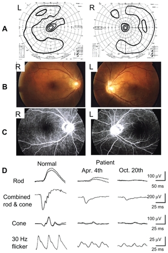
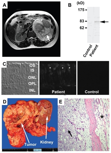
Figure 2 Systemic and histologic findings in a case of paraneoplastic retinopathy. A) Abdominal MRI showing a large retroperitoneal mass (arrow), which compressed the left kidney. B) Western blot analysis of patient’s serum using bovine retinal protein. The serum reacted to an 83 kD antigen (arrow). C) Immunohistochemical analysis using patient’s serum demonstrates autoreactivity against the photoreceptors of bovine retina. D) Gross appearance of tumor. E) Microscopic appearance of retroperitoneal tumor (× 20). Two characteristic patterns of well differentiated liposarcoma (asterisk) and dedifferentiated fibrotic sarcomatoid tissue (arrow) can be seen.