Figures & data
Figure 1 Extensive eyelid swelling combined with less obvious swelling of the temples and cheeks.
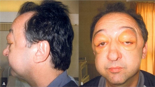
Figure 2 Axial A) and coronal B) contrast-enhanced fat-suppressed T1-weighted MR images showing bilateral eyelid, lateral rectus oculomotor, masticatory and temporal muscle enlargement and enhancement.
Abbreviation: MR, magnetic resonance.
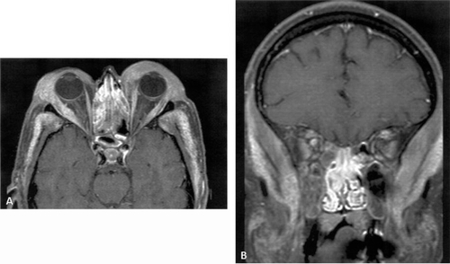
Figure 3 Diffuse infiltrate rich in lymphocytes, foamy histiocytes, and giant cells (hematoxylin and eosin: x250). A) Giant cells of Touton type, B) nodular lymphoid infiltrate.
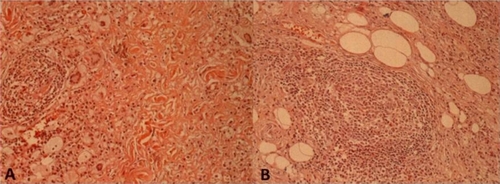
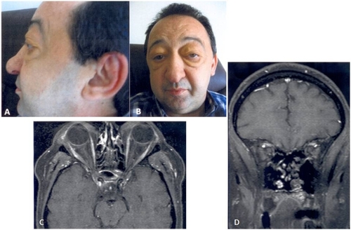
Figure 4 Six months of treatment. Significant regression of eyelid swelling (A, B). Follow-up MRI: axial (C) and coronal (D) contrast-enhanced fat-suppressed T1-weighted MR images shows normalization of eyelid, lateral rectus oculomotor, masticatory and temporal muscle.
Abbreviation: MRI, magnetic resonance imaging.