Figures & data
Figure 1 The ECG for case one demonstrated prominent ST elevations in the anterior and inferior leads.
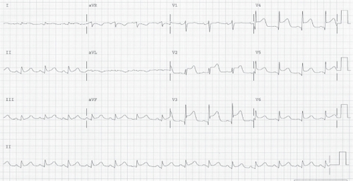
Figure 2 In case one, noncritical, distal lesions of the right coronary artery (RCA) (a) and noncritical atherosclerotic lesions of the left main coronary artery (b) did not correspond with the wall motion abnormality observed on ventriculography. In case two, there was no evidence of coronary arterial stenosis of either the RCA (c) or the left anterior descending artery (d).
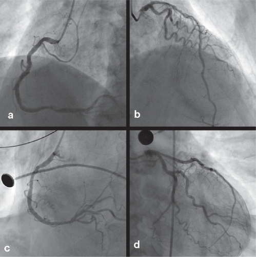
Figure 3 In the second case, the ECG was notable for new Q-waves in inferior and precordial leads as well as ST elevation suggestive of acute ischemia or injury in the anterior-septal leads.
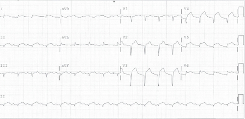
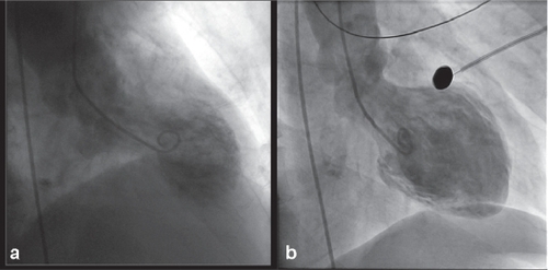
Figure 4 (a) This image is a left ventriculogram obtained during systole in the first patient. Hypercontractility of the basal segment and akinesis of the apical segment create the apical ballooning effect. (b) The ventriculogram obtained in the second case also demonstrates apical ballooning.