Figures & data
Figure 1 The organization of apoB100 on the LDL particle. Two of the sites (A and B) involved in the binding of apoB100 (and LDL) to proteoglycans are indicated in the figure and their primary sequence is given below. Site B is also the binding site for the LDL receptor.
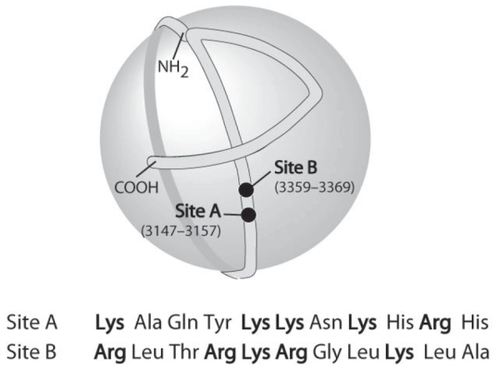
Figure 2 The assembly of VLDL. During its biosynthesis, apoB100 (1) is translocated to the lumen of the endoplasmic reticulum and lipidated by MTP to form a pre-VLDL particle (2). Pre-VLDL is further lipidated to form VLDL2 (5). Alternatively, pre-VLDL and misfolded apoB100 (3) can be retained and degraded in the cell (4). The VLDL2 is transferred to the Golgi apparatus (6, 7) and is either secreted or further lipidated to form VLDL1 (8). Fatty acids are released from cytosolic lipid droplets and used for the formation of triglycerides, which are assembled into VLDL (8).
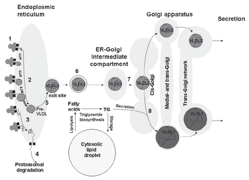
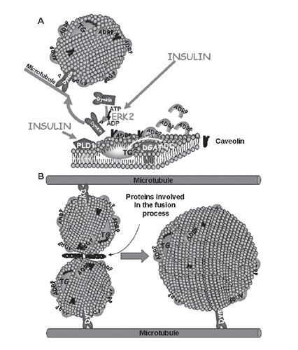
Figure 3 (A) Lipid droplets are formed as primordial droplets at the microsomal membranes. Insulin stimulates the droplet formation by activating PLD1 and ERK2. ERK2 phosphorylates the motor protein dynein, which is then recruited to the droplets to promote their formation and fusion. Diacylglycerol acyltransferase (DGAT) catalyzes the formation of triglycerides (TG) from diglycerides (DG) and fatty acids (FA). It has been suggested that the lipid droplet is initiated by the oiling out of triglycerides between the leaflets of the membrane. (B) Primordial droplets increase in size by fusion. This process depends on dynein and its interaction with microtubules. Moreover it involves specific proteins that catalyze fusion between the droplets.