Figures & data
Figure 1 The effect of ischemic preconditioning on infarct size after ischemia/reperfusion. Upper panel:infarct size expressed as percent of risk zone size in control and preconditioned (IPC) rabbit hearts (from CitationIliodromitis et al 2006); lower panel: the absolute infarct volume plotted against absolute risk zone volume from control (closed symbols) and preconditioned (open symbols) hearts (from CitationIliodromitis et al 2004).
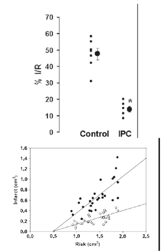
Figure 2 Schematic representation of the death receptor (extrinsic) pathway of caspase activation to provoke the mechanism of apoptosis.
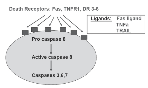
Figure 3 Schematic representation of the mitochondrial death (intrinsic) pathway leading to the formation of apoptosome, activation of caspases and apoptosis.
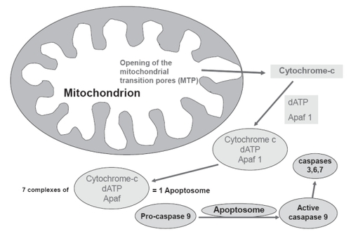
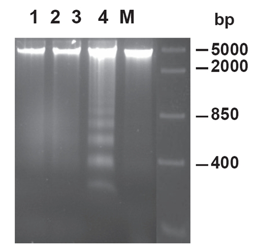
Figure 4 Effect of ischemic preconditioning on DNA fragmentation in rabbit hearts subjected to ischemia/reperfusion Lane 1 represents control non-ischemic tissue; lane 2 represents ischemic tissue after ischemia without reperfusion; lanes 3 and 4 represent ischemic tissue after ischemia/reperfusion in nonpreconditioned and preconditioned hearts, respectively. M, marker lane (from CitationLazou et al 2006).