Figures & data
Figure 1 Volume parameters obtained by using magnetic resonance imaging before (crosshatched bars) and after CABG (hatched bars). Data are mean ± SEM. ESV, Endsystolic volume; EDV, Enddiastolic volume. Numerical values above error bars indicate level of statistical significance between the groups (Student’s t-test for unpaired samples).
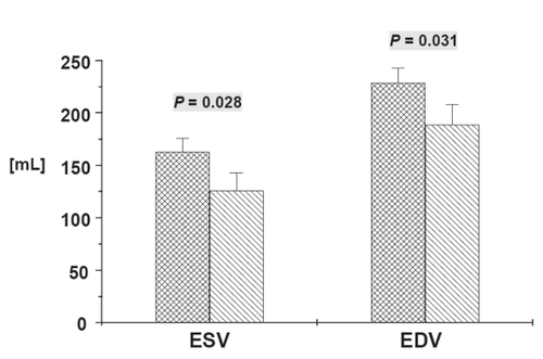
Figure 2 Left ventricular ejection fraction (LVEF) obtained by using magnetic resonance imaging before and after surgery. Data are mean ± SEM. Numerical value above error bar indicates level of statistical significance between groups (Student’s t-test for unpaired samples).

Figure 3 MR Imaging of a 64 year old male before (top line) and after (bottom line) 3-fold CABG. SSFP Cine images in enddiastole (A) and endsystole (B) reveal the severely impaired global LV function before surgery (EF 30%) with akinesia in the apical inferoseptal and anterolateral wall and the apex (segments 14, 16, and 17) and hypokinesie in the basal and mid-ventricular lateral wall. The contrast-enhanced TurboFLASH image (C) shows broad subendocardial late enhancement (bright signal) in the apical septum, thin LE in the lateral wall and transmural LE in the apex meaning chronic scar. LV function after surgery (D, E) shows no improvement in the apical septum and the apex, whereas the complete lateral wall improved and became normokinetic. No changes in scar extent (F). Global LV function improved to EF 40%, LV volumes decreased.
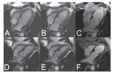
Table 1 Preoperative patient data
Table 2 Intra- and postoperative patient data