Figures & data
Table 5 Cross reactive intralesional T-cell clones from RHD patients
Table 1 Clinical data of RHD patients
Table 2 Echocardiography data before surgery and until seven days post surgical treatment
Figure 1 Kaplan-Meier survival curve: follow-up of 25 rheumatic valve disease patients. (A) Event-free survival (death and reoperation); (B) probability of survival.
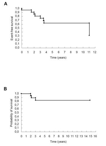
Table 3 Histopathological data from valvar and myocardial tissues of rheumatic heart disease patients
Table 4 Evaluation of heart-tissue infiltrating lymphocytes subsets
Figure 2 The presence of heart tissue T lymphocytes is related with rheumatic activity. The number of infiltrating T and B lymphocytes and their relation with rheumatic activity in both valve and myocardium tissues are presented. Black bars, mean tissue with rheumatic activity and white bars, without rheumatic activity. *P = 0.03; **P = 0.03. HPF, high power microscopic field.
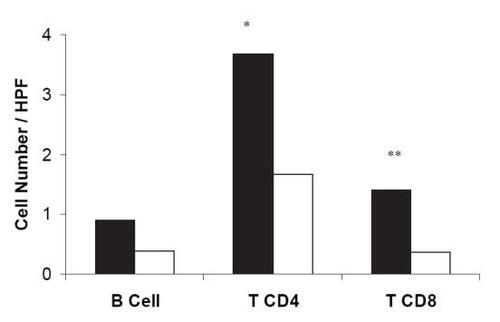
Figure 3 Heart infiltrating T-cells. Heart tissue biopsies were cut in small fragments and kept in culture until establishment of T-cell lines. The picture represents lymphoblasts cultivated for 15 days in Dulbecco’s modified Eagle medium supplemented with IL-2. Photograph from Immunology Laboratory, Heart Institute; original magnification ×200.
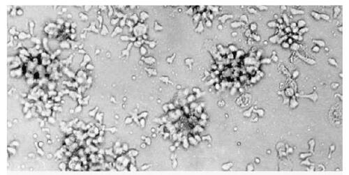
Table 6 Peripheral blood reactivity against streptococcal M5 peptides and cardiac proteins