Figures & data
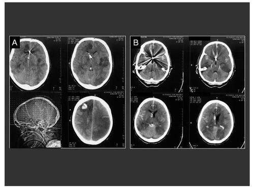
Figure 1 Upper sequence: SAH in a 48 year-old patient, H&H 4; (A) Preoperative CT-scan showing a right temporal mass occupying bleeding and secondary brain swelling; (B) Preoperative antero-posterior cerebral angiography of the right internal carotid artery demonstrating a multilobular giant aneurysm on the middle cerebral artery (white arrow) with vascular displacement; (C) Postoperative angiographic control showing a complete aneurysm occlusion and the borders of the decompressive craniotomy (black arrow); (D) Postoperative CT-scan demonstrating the clot-removal and the surgical decompression. Lower sequence: Fifty nine year-old woman with massive subdural and SAH and clinical herniation signs, H&H 5; A) Preoperative CT-scan demonstrating the right hemispheric subdural hemorrhage and the acute brain shift of the midline structures; (B) CT-angiography displaying the aneurysm (arrow) arising from the internal carotid artery; (C) Postoperative CT-scan demonstrating the brain reexpansion the surgical decompression.
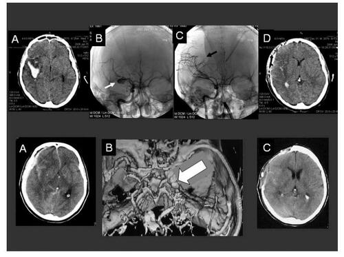
Figure 2 Eleven year-old child with H&H 5 SAH and clinical herniation signs; (A) Preoperative CT-scans demonstrating a left frontal mass occupying bleeding with intraventricular dissemination and acute hydrocephalus; (B) Postoperative CT-scans confirming the clot removal and the placement of the ventricular drainage. The borders of the decompressive craniotomy are well defined; (C) Preoperative cerebral angiography of the left internal carotid artery demonstrating the aneurysm (white arrow) and the caliber narrowing of the anterior cerebral artery (black arrow); (D) Postoperative, 10 hours later performed control angiography confirming the occlusion of the aneurysm and the improved cerebral perfusion.
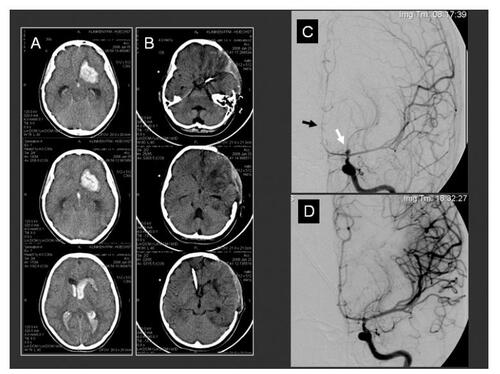
Figure 3 Two endovascular treated patients with delayed secondary ischemic lesions. These patients were both grade V at admission and with an acute CSF obstruction in CT-scans. Ventriculostomy, angiography and aneurysm coiling were in both patients performed. Multiple intracerebral hypodensities and the coils artifacts can be observed in both cases. (A) CT-scan control 5 days after aneurysm coiling and before decompressive procedure in this 44-years-old patient showing two coiled (AcoA and pericallosa) aneurysms. ICP values were obtained from the right side placed intraventricular catheter; (B) CT-scan control 4 days after aneurysm coiling (AcoA) and before decompressive procedure in a 63-years-old patient showing similar findings than patient A. Both cases underwent prolonged endovascular procedures.