Figures & data
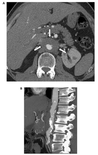
Figure 1 CT angiography of the aorta performed with 64-row MDCT. (A) Axial image at the level of the renal arteries showing narrowing of the aortic lumen, aortic wall thickening and periaortic rind (arrows). (B) Multiplanar reformat image showing periaortic thickening confined to the abdominal aorta (A) (dashed line). The thoracic aorta is spared. Numerous calcified plaques are seen mainly in the infrarenal segment of the abdominal aorta.