Figures & data
Figure 1 Layers of aortic wall.
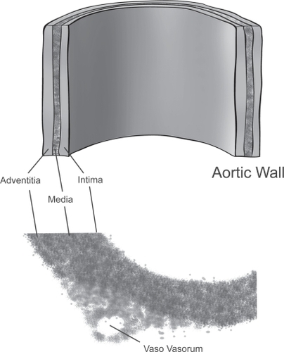
Figure 2 Aortic dissection. Actual specimen (A) and intravascular imaging (B) show both the true and false lumen.
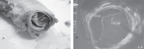
Table 1 Characteristics of patients (N = 951) with acute aortic dissection – from the International Registry of Acute Aortic DissectionCitation8
Figure 3A Debakey classification.
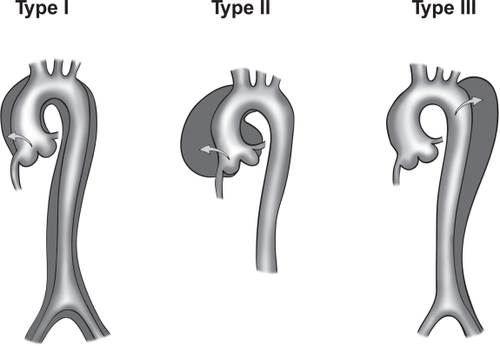
Figure 3B Stanford classification of aortic dissection. Stanford type A includes dissections that involve the ascending aorta, arch, and descending thoracic aorta. Stanford type B includes dissections that originate in the descending (and thoracoabdominal) aorta, regardless of any retrograde involvement of the arch.
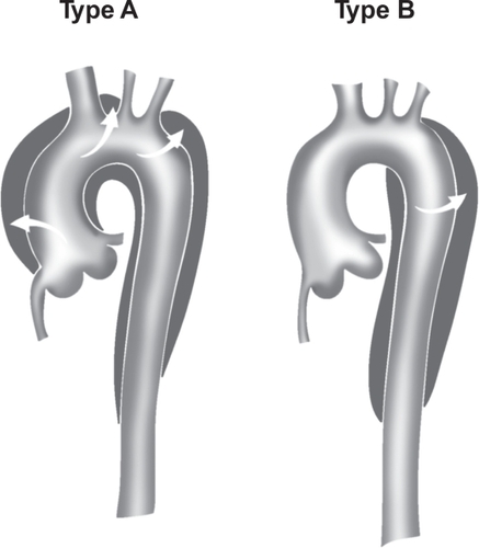
Figure 4 Computer tomography with enhanced contrast showing type B dissection.
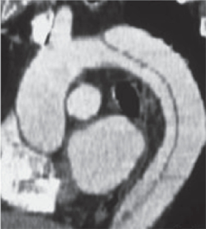
Figure 5 Intravascular imaging of aortic dissection.
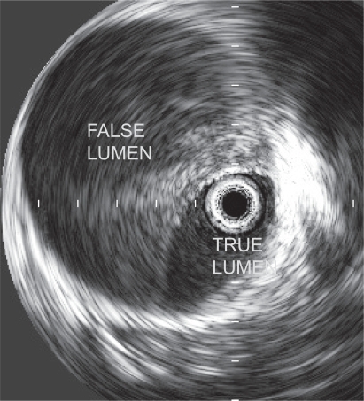
Table 2 Approach to acute aortic dissections (AAD) in the emergency department
Table 3 Indications for endovascular or surgical intervention in patients with type B AAD