Figures & data
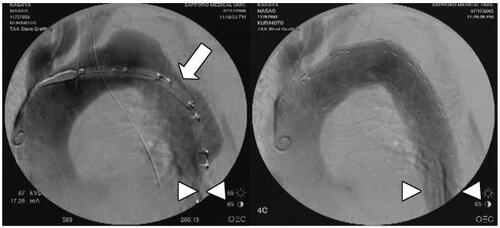
Figure 1 92-year-old man with distal aortic arch aneurysm ruptured into mediastinum. The left subclavian artery is covered by the stent graft, but full patency of the left carotid artery is achieved by the fenestrated stent graft (white arrow).
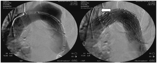
Figure 2 74-year-old woman with blunt aortic injury associated with intracranial hemorrhage and pelvic fracture. Emergency stent-grafting was performed 2 hours after arrival. Typical aortic isthmus injury (white arrow) is well excluded by the stent graft.
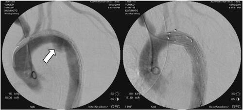
Figure 3 57-year-old man with acute type IIIa aortic dissection ruptured into the left pleural cavity. In addition to complete entry closure, full patency of the left carotid artery and the left subclavian artery is achieved by the fenestrated stent graft (white arrow).
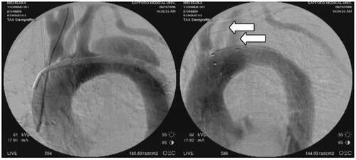
Figure 4 81-year-old man with acute type IIIa aortic dissection complicated with visceral and leg ischemia. The entry site (white arrow) was revealed by intravascular ultrasound (IVUS) before deployment of the stent graft. Critically compressed true lumen of the distal descending thoracic aorta is sufficiently expanded by the entry closure using the stent graft (white arrowheads).