Figures & data
Figure 1 Fundus photographs before and after pioglitazone treatment. A and B: 4 months before pioglitazone treatment, macula edema is not present in either eye (A shows the right eye, B the left eye). C and D: during pioglitazone, DME is present in both eyes but it is difficult to detect in fundus photographs because the DME is very diffuse (C shows the right eye, D the left eye).
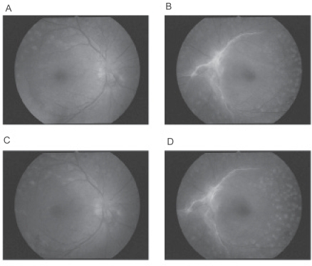
Figure 2 Fluorescein angiograms before and after pioglitazone treatment. A and B: 6 months before pioglitazone treatment, neovascular abnormalities with no leakage are present in both maculas but the activity is low and no DME is observed (A shows the right eye, B the left eye). C and D: During pioglitazone treatment, the status of proliferative diabetic retinopathy is not changed before and after pioglitazone treatment. DME is difficult to detect in the fluorescein angiograms because it is very diffuse. Optical coherence tomography shows the diffuse DME more clearly (C shows the right eye, D the left eye).
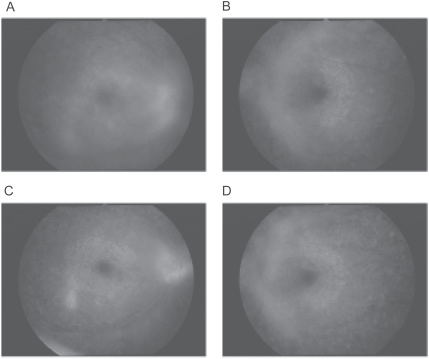
Figure 3 OCTs before and after pioglitazone treatment. A: 3 months before pioglitazone treatment, macular edema is not present in both eyes. B: 2 weeks after pioglitazone, severe diabetic macular edema can be seen in both eyes. C: 6weeks after pioglitazone, DME is worse and a serous retinal detachment is present in the right eye. D: 2 weeks after cessation of pioglitazone, visual acuity has improved but severe DME is still present in the OCT images of the right eye. E: 2 months later after receiving a half-dose of spironolactone, DME is significantly reduced in both eyes.
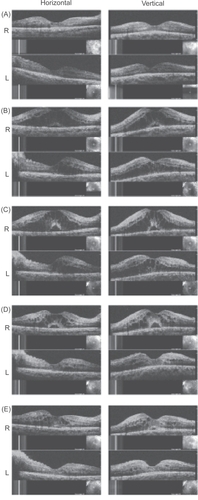
Table 1 Clinical course of systemic condition