Figures & data
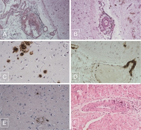
Figure 1 A) Association of lymphocytic angiitis and amyloid deposits infiltrating the wall of leptomeningeal vessel and perivascular cuffing of mononuclear cells (hematoxylin-eosin-safran original magnification X 10). B) Mononulear cells surrounding a non amyloid cortical vessel with severe intimal fibrosis occluding the lumen (hematoxylin-eosin-safran original magnification X 20). C) Amyloid deposits present in the senile plaques immunostained for bA4 (original magnification X 40). D) Amyloid deposits present in the wall of vessels in the neocortex immunostained for bA4 (original magnification X 40). E) Neurofibrillary tangles immunostained for tau protein (original magnification X 10). F) Giant cell arteritis in the myocardium (hematoxylin-eosin-safran original magnification X 40).